DO TWO TYPES OF CELLS EXIST IN POPULATION OF
HUMAN ERYTHROCITES?
Rudenko S.V.1 , Mohammed Khaleel Saeed2
1Institute of Cryobiology and Cryomedicine
Problems, NASU, 23, Pereyaslyavskaya str., Kharkov, 61015, Ukraine, e-mail:
rsv@kharkov.ua
2V. N. Karazin Kharkov National University,
4 Svobody aq., Kharkov 61022, Ukraine, e-mail: mohkh66@gmail.com
ABSTRACT
This work characterizes the conditions under which discoid population of red blood cells is separated into sub-populations of coexisting discocytes, echinocytes and stomatocytes in various proportions. The fact that, an existence of mixed population containing approximately equal amount of echinocytes and stomatocytes is possible indicates that, cell population contains at least two types of cells responding to the shape in opposite manner, when external condition are changed. While echinocytes and stomatocytes are considered as opposite classes of cell shape with negative and positive curvature, their simultaneous coexistence can not be explained on the basis of modern concepts of red blood cell shape regulation, thus this points to need a more complex description of these phenomena.
Key words: erythrocyte, shape, morphological response, sucrose, benzolkonium
chloride, bilayer couple theory.
INTRODUCTION
A population of human erythrocytes is functionally specialized by cells, which differ in size, surface area, concentration, hemoglobin amount and many other physiological and physical parameters [1]. At normal physiological conditions, erythrocytes have shape of biconcave disk and relatively small coefficient of variation of basic cellular parameters that can be regarded as a fairly homogeneous population of cells. In this sense, a form of erythrocytes has the smallest distribution, as other normal forms, except biconcave disk are extremely rare. Homogeneity implies that the changes in conditions will react the same way on all cells. For example, by decreasing medium tonicity, all cells swell, while increasing the tonicity they compressed. After echinocyte factor influence, all the cells become echinocytes, and after stomatocyte - stomatocyte. Under the action of hemolytic factors of all cells undergo a certain degree of hemolysis. The difference in the effect on the individual cell is only in degree but not direction, of change, which is determined by its cellular characteristics. The homogeneity of the population of red blood cells and is performed in the phenomenon of morphological response - three-phase changes in the shape of cells when they are placed in non-electrolyte medium of sucrose, which occur very synchronously [2,3]. The purpose of this study was to characterize the conditions under which homogeneity may be disrupted and lead to opposite cellular responses in terms of the morphology of red blood cells, which appear at different stages of morphological response.
METHODS OF INVESTIGATION
All the experiments were conducted on fresh human erythrocytes, which were kindly presented by clinical laboratory of the State Institution Institute of Medical Radiology named by S.P. Grigoriev of Academy of Sciences of Ukraine. Blood was taken into vacuum tubes containing EDTA as an anticoagulant. After the spontaneous deposition of sediment of 0.15 ml, solution was washed once in saline (150 mM of NaCl) and once in isotonic HBS (150 mM NaCl, 5 mM HEPES, pH 7.4). The resulting precipitate is diluted, leading to a final volume of 1 ml and used as stock-suspension.
To study the dynamics of morphological response (MR) cells in the standard sucrose medium (SSM) (0,3 M of sucrose, pH 5,8-6,2) used a dual-formmeter aggregometer FA-01. Terms and conditions for procedure measurements are described in [4].
Depletion of red blood cells by ATP was carried out by keeping the drain-cell suspension at 37 ° C for 24 hours. The control sample of the same cells in parallel was kept at +2 ° C. To fix the form of red blood cells in 2 ml of cell suspension was added 0.2 ml of fixing agents of the following composition: FRa - 5% glutaraldehyde, 20 mM phosphate buffer, 150 mM NaCl, pH = 6.4; FRb - 4% formalin, 50 mM phosphate buffer, pH = 7,4; FRab - a mixture of reagents FRa and FRb in a 1:1 ratio. Once the cells were kept in this solution for several hours, the samples were centrifuged, the sediments were studied microscopically, and photographed with an increase in 800. For this purpose was used the stock detergent solution Benzalkonium chloride (BzA) «Fluka» (Germany) at a concentration of 0.4 mg/ml and ophthalmic drug Timolol (active substance Timolol, 5 mg/ml) "Farmak" (Ukraine). Experiments were performed at room temperature 22-24 ° C.
RESULTS AND DISCUSSION
In the sucrose solution, cells dynamically change their shape, undergoing three-phase morphological response (MR), in which the first phase is echinocyte, the second is discocyte, and third is stomatocyte [2,3]. Fig. 1 compares the MR of control and ATP-depleted red blood cells and the effect of fixing reagents were added to its maximum and at the end of phase 3. It is seen that the fixation of the two agents of control of red blood cells does not lead to significant changes in the shape index peak, which indicates its good value at these points. MR of ATP depleted red blood cell is different from the control so that it is more tightened over time and has a lower value of shape index at the maximum.
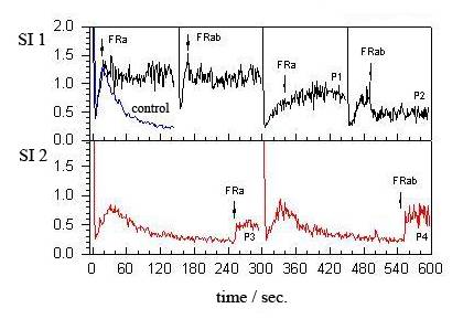
Fig. 1. Changes of shape index(SI) in the native and depleted on ATP erythrocytes in the course of morphological response in the standard sucrose medium (SSM) and under the effect of the fixing reagents at its various stages. Arrows and notations near them designate the type of the added reagent. Data for the native cells are cited on [SI]1, curves 1 and 2, remaining curves relate to ATP depleted erythrocytes. Legend FR means adding 0.2 ml of the fixing reagents to 2 ml of erythrocyte suspension.
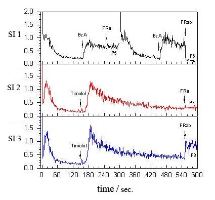
Fig. 2. Changes of Index form (SI) in the native erythrocytes in the course of morphological response in an isotonic medium mannitol (0.3 M mannitol) Index form (SI) and SSM (SI 2 , SI 3), the action BzA, Timolol and fixing reagents at its various stages. Arrows and notations near them designate the type of the added reagent, as in Fig. 1.
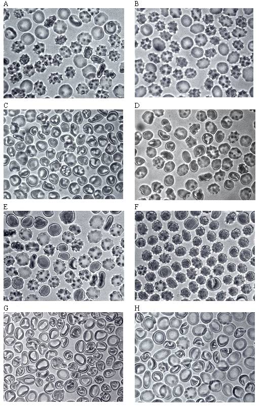
Fig. 3. The morphology of red blood cells after fixation at various time points of morphological response, the corresponding data presented in Fig. 1 and 2. Each picture corresponds to the fixation point P1 to P8.
А – Р1; B – Р2; C – Р3; D – Р4; E – Р5; F – Р6; G – Р7; H – Р8;
This suggests that these red cells do not fully restore the discoid shape, as compared with native cells, and in addition, the reagent FRab itself influence on the shape of cells, reducing the number of shape index. When adding latches to phase 3, an increase in shape index, which indicates a greater influence on the shape of fixers themselves. These microscopy data is shown on Fig. 3, the population of ATP-depleted cells at the peak represented a mixture of discocytes, echinocytes and, in contrast to control cells, which are mainly represented by discocytes having a high value of shape index (data not shown). In phase 3 these cells after fixation FRa have a form of stomatocyte that is consistent with earlier data for native cells [2], but when commiting to the same point in time reagent FRab population is divided into echinocytes and stomatocytes. Figure 2 shows that after the action of BzA mannitol environment fixation both agents leads to echinocytes and stomatocytes, echinocytes are in major after FRab fixation. In saccharine environment, after the second wave of MR, induced by Timolol, stomatocytes formed by fixing FRa, and mixed population stomatocytes discocytes and after fixing under the same conditions. Thus, these data show that in the MR, according to environmental conditions and phases of the process, after the action of fixing reagents may be formed discocytes, a mix of discocytes echinocytes, discocytes and stomatocytes and stomatocytes and echinocytes. It is interesting to note that often, as shown in Fig. 3, the ratio of these forms is approximately one to one. The observed phenomenon is not limited to the terms set forth above. Because many substances are able to modify the standard MR in sucrose medium, affecting its various phases [2,3] the division of cells in subpopulation expressed there and in other situations. The variety and diversity of conditions determines the response of cellular reactions, but at present it is difficult to identify some regularity in the appearance and orientation of the division of forms of cells in the population. This is due to the fact that, depending on the ionic composition of the medium and component fixing reagents exhibit a echinocytes then stomatocytes type of action and then more, then at least influence the current shape of cells during fixation. What is the reason remains unclear, and only by experiment, and choose conditions, such as fixing a few of the reagents can be approached in a more or less adequate fixation.
Our data shows that the level of ATP in the cell affects the dynamics of its MR. A lower value of shape index at the peak may be due either to the fact that some of these cells are not able to recover discoid shape in the MR, and with the resynchronization of the cell population where all cells are discoid but at different times. As a result, in this case, we always have a mixture of different shapes, which gives a low value of shape index [4]. This hypothesis suggests that the peak width of MR for ATP-depleted cells two times greater than for controls. If so, this suggests that the ATP-depleted cells are also able to demonstrate normal MR. On the other hand, it is possible that these cells make a direct transition from echinocytes in stomatocytes, bypassing the discoid shape, which has been shown in other conditions [5]. We are observing the separation of erythrocytes subpopulations when fixing reagents, which are in themselves also cause changes in cell shape. Consequently, the division is the final result of the influence of the ion source and environment of the striker. Whether such a division in the population does the same when it is no catches is still unclear.
We believe that our findings, especially the division of the population in echinocytes stomatocytes without the presence of discoid forms, are essential, as it is now difficult to suggest some reasonable scheme for their explanation. The existing model for control of shape of red blood cells [6-13], ultimately boil down to the original hypothesis put forward by Hoffmann [6], Sheets and Singer [7], who proposed that the mechanism involves an imbalance membrane and small changes in surface area between the ΔA0 bilayers of the cell membrane (bilayer couple theory) and differ in what kind of structure - the lipid bilayer, membrane cytoskeleton, or both, or the conformation of transmembrane proteins play a key role in the generation of the ΔA0. None of these models does not allow for the possibility that the same conditions, some cells will turn into echinocytes, and another part in stomatocytes that observed in our experiments.
To explain the obtained data suggest two hypotheses. The first is that the population of human erythrocytes is not conditionally uniform, as is commonly believed, but contains at least two cell types - e-RBC, who tend to echinocytes and s-RBC, who tend to stomatocytes. Different reactivity of these cell subtypes, possibly genetically caused, and determines that under certain condition some cells move toward to stomatocytes and others in the direction of echinocytes. This suggests that the mechanisms of regulation of these subtypes form must differ. If we assume that the bilayer couple theory is basically correct, the difference mechanisms could consist in the fact that the factor leading to an increase in ΔA0 in some cells, leads to a decrease in ΔA0. This can be realized due to differences in the sensory system that transmits information from the stimulus to the executive system that controls the ΔA0. Another hypothesis is that cells are still the same, but depending on the environment may be, at least in two states S and E, which determine their reactivity. Being similar, these two hypotheses are fundamentally different in that if the first e-RBC and s-RBC are independent of cell types that can not pass each other, then the second, any red blood cell, under certain circumstances, may be as e and in the cell. Recently, we described several states form red blood cells, which arise in the course of the morphological response, and showed that between them may be different transitions [14]. In this light, the second hypothesis seems more plausible, but more research is needed to identify factors that control the transitions of cells in different states, and their role in the physiology of red blood cells.
The authors thanks to the State Institute of Medical
Radiology named by S.P. Grigorieva and to AMS of Ukraine for their support and
providing a base for this work.
REFERENCES
1. Investigation of blood sytem in clinicals.
(1997). Eds., Kozinets, H. I and Makarova, V. A Edition. Moscow: Triada-X., 480.
2. Rudenko, S. V. (2009) Blood Cells Mol. Dis., 42, 252-261.
3.
Rudenko, S. V. (2009) Bioelectrochemistry.,
75, 19-25.
4. Rudenko, S. V, Krouf, J. H and Tablin, F. (1998) Biochemistry,
63, 46–55.
5. Rudenko, S. V., Saeed, М. Кh. (2010) Biochemistry. Moscow: 75, (N8), 1025-1031.
6. Hoffman, J. F. (1972) Nouv.
Rev. Fr. Hematol., 12, 771-774.
7. Sheetz, M. P and Singer, S. J. (1974) Proc. Natl. Acad. Sci.U.S.A, 71,
4457-4461.
8. Lim, H.W.G., Wortis, M. and Mukhopadhyay, R. (2002) Proc. Natl. Acad. Sci. U.S.A, 99, 16766-16769.
9. Mukhopadhyay, R, Lim, H.W.G. and Wortis, M. (2002) Biophys. J., 82, 1756-1772.
10. Wong, P. (1999) J. Theor. Biol., 196, 343-61.
11. Sheetz, M. P. and Singer, S.J. (1976) J. Cell Biol., 70,
247-251.
12. Tachev, K. D., Danov, K. D. and Kralchevsky, P. A. (2004) Colloids Surf. B. Biointerfaces, 34, 123–140.
13. Gimsa, J and Ried, C. (1995) Mol.
Membr. Biol., 12, 247-254.
14.
Rudenko, S. V. (2010) Biochim. Biophys. Acta, 1798,
1767-1778.
DO TWO TYPES OF CELLS EXIST IN POPULATION
OF HUMAN ERYTHROCITES?
S.V. Rudenko1,
Mohammed Khaleel Saeed 2
1Institute for
Problem of Cryobiology and Cryomedicine of NASU, 23 Pereyaslavskaya St.,
Kharkov, 61015, Ukraine; e-mail; rsv@kharkov.ua
2V.N.Karazin Kharkov
National University, 4 Svobody Sq., Kharkov, 61022, Ukraine, e-mail: mohkh66@gmail.com
This work characterises the conditions under which the discoid population of red blood cells is separated into subpopulations of coexisting discocytes, echinocytes and stomatocytes in various proportions. The fact, that an existence of mixed population containing approximately equal amounts of echinocytes and stomatocytes is possible indicates that cell population contains at least two types of cells responding by shape in opposite manner when external conditions are changed. While echinocytes and stomatocytes are considered as opposite classes of cell shape with negative and positive curvature their simultaneous coexistence can not be explained on the basis of modern concepts of red blood cell shape regulation, thus points to need a more complex description of these phenomena.
Key words: erythrocyte,
shape, morphological response, sucrose, benzalkonium chloride, bilayer couple
theory.