Synthesis of composite based on vinyl ether of ethylene glycol structured nano particles hydroxyapatite
Temirkhanova G.E., Trubitsin
M.A., Mun G.A., Urkymbaeva P.Y
National research university
Belgorod State University, Belgorod
Al-Farabi Kazakh
National University, Almaty
Abstract:Millions of
people are suffering from bone defect arising from trauma, tumor or bone diseases.
Therefore, there is a growing need for the development of biocomposites with
excellent bioactivity and compatibility. In this study,hydroxyapatite (HAp)
nanorod embedded composite was prepared using vinyl etherof
ethylene glycol(VEEG) as a matrix.The role of VEEG
composition on the crystallite size, degree of crystallinity, functional groups
and morphology of nanocomposites were characterised by TEM analysis. The
results indicated that the size and crystallinity of Hapnano particles decreases
with increase in VEEG concentration in the composite. This shows the size
control effect of VEEG concentration on HApnanorods. Due to the chemical bond
interactions between HAp and VEEG. TEM micrograph confirms the presence of
Hapnano rod crystals in VEEG matrix.
1.Introduction
The calcium phosphate based bioceramics
particularly hydroxyapatite (HAp) play an excellent role in biomedical
applications owing to their excellent biocompatible, osteoconductive and
bioactive properties, and its close chemical and physical resemblance to
mineral component of bone tissue, enamel and dentin. The major mineral phase of
bone is hydroxyapatite (HAp) with a ratio of calcium-to-phosphate is 1.67 which
is embedded as nanocrystalline form in collagen triple helix structure.
Currently, researchers are trying to mimic this natural nano composite system
for tissue engineering applications. Since, the nanoHAp with high surface area
to volume ratio is more desirable to increase their contribution in bone/tooth
implants, adsorbents, gene delivery and immune sensor. However, the brittleness
and poor performance of mechanical stability of pure HAp limit its use for the
regeneration of non-load-bearing bone defects and tissue engineering
applications.Composite biomaterials like metal and polymer matrix are used to
improve the mechanical compatibility of nanoHAp (n-HAp). Generally, the
composite biomaterials are prepared by using biocompatible/biodegradable
synthetic/natural polymers [1].
The
inorganic minerals such as hydroxyapatite, bioactive glasses, metal oxides, and
carbon nanotube are incorporated into polymer matrixes to impart bioactivity.
This enables us to developed the composite with desired properties. The
addition of nanosized particles is desirable to develop the composite with a
good mechanical strength since the natural bone contains mineral crystals which
are at the nanometer scale and embedded in the collagen matrix. The polymer
composites are designed to meet the specific requirement of biomedical
applications like tissue engineering and drug delivery system. The right choice
of the composition of both filler and polymer matrix are essential in addition
to the process method to obtain suitable biopolymer composites. Recently,
attempts have been made to develop nanocomposites, wherein n-Happarticles are
embedded in VEEG polymeric matrices [2].
An
extensive study have been made on both natural (collagen, gelatin, silk
fibroin) and synthetic (polyethylene, polyamide, chitosan, polystyrene, poly
(vinyl alcohol) and polyetherethilenglicole) polymers to overcome the
mechanical problems associated with bioceramics in bone tissueengineering
applications [3-5].Among the above polymers, VEEG remain one of the widely used
polymer group of biomaterials applied for medical implants. This usage is due
to its segmented block co-polymer character. This wide range of versatilityin
terms of tailoring their applications such as tissue scaffolding, artificial
cartilage and biodegradable scaffolds.
With
the superior combination of the synergic effect and biocompatible HAp and the
adjustable biodegradability of polymer matrix, HApnanorod embedded VEEG
composites were prepared under controlled environment. The obtained
nanoHAp/VEEG composites were characterised in light of crystallite size, degree
of crystallinity, morphology, biological and mechanical properties [6].
2.
Formation mechanism of HAp/VEEGnanocomposite
Fig. 1 shows the schematic representation
of the synthesis of HAp/VEEG nanocomposite. When the calcium hydroxide was
added to the VEEG solution, the Ca2+ ions were attached with OH-group
in the VEEG matrix. Following the above step, orthophosphoric acid was added
drop by drop into the above mixed solution. As a result, PO3-ions
bind to the –OH- and Ca2+group to form hydroxyapatite
particles and the VEEG matrix regulates the growth of c-axis of HApnanorod.
|
|
|
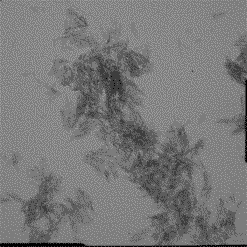
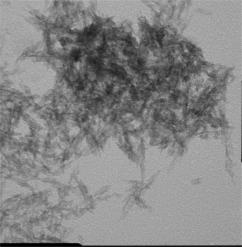
Figure 1.TEM micrographs of the prepared
nanohydroxyapatite samples: (a) without polymer, (b) with polymer
3.Transmission electron microscopy analysis
TEM(Jeol
JEM-2100) images of pure n-HApand vinyl
etherof ethylenglycolcompositions are illustrated in Fig.1. The
TEM picture shows that particles exhibit nanorod morphology. In case of
composites, when the composition of VEEG is added to HAp, the rod-like
morphology starts to disappear. According to TEM analysis, the particles are
homogeneously dispersed in polymer matrix. Further, the micrograph does not
show any notable indication for the existence of agglomeration.
4. Conclusion
In
the present work, novel hydroxyapatite/vinyl
etherof ethylenglycolnanocomposite is prepared by simple chemical
route. It inferred that the composition of VEEG shows significant influence on
particle size, degree of crystallinity and microhardness, which facilitate to
optimize the composition of composite for particular applications.
Reference:
1. M. Li, X.
Xiao, R. Liu, C. Chen, and L. Huang. Structural characterization of
zinc-substituted hydroxyapatite prepared by hydrothermal method, J. Mater.Sci:
Mater. Med. 19: 797–103 (2008).
2. S. Bose, and S. K. Saha. Synthesis and
characterization of hydroxyapatite nanopowders by emulsion technique, Chem.
Mater. 15:4464-4469 (2003).
3. Y. Ding, J. Liu, H. Wang, G. Shen, and R.
Yu.Apiezoelectricimmunosensor for the detection of α-fetoprotein using an
interface ofgold/hydroxyapatite hybrid nanomaterial, Biomaterials 28: 2147–2154
(2007).
4. H. Wang, Y. Li, Y. Zuo, J. Li, S. Ma, and L.
Cheng. Biocompatibility and osteogenesis of biomimetic
nanohydroxyapatite/polyamide composite scaffolds for bone tissue engineering,
Biomaterials 28: 3338–3348 (2007).
5. V. S. Komlev, S. M. Barinov, and F.
Rustichelli.Strength enhancement of porous hydroxyapatite ceramics by
polymerimpregnation, J. Mater. Sci. Lett. 22: 1215–1217 (2003).
6. N. MeenakshiSundaram, E. K. Girija, M.
Ashok, T. K. Anee, R. Vani, and R. Suganthi.Crystallisation of
hydroxyapatitenanocrystals under magnetic field, Mater. Lett. 60: 761-765
(2006).