Kolesnyk Vladimyr
Bukovinian State Medical University, (Chernivtsi) Ukraine
MORPHOLOGICAL PATTERNS OF THE VASCULAR PLEXUS OF LATERAL VENTRICLES OF
THE CEREBRUM
The strides made
by modern neuroanatomy, neurosurgery, an increase of surgical interferences in
the region of the cerebral ventricles and on the vascular plexuses,
experimental-clinical investigations that are carried out on these particular
formations and attemps of their transplantation (Skinner S.L. et al.,
2006-2009; Matsumoto N. et al., Thanos C.G. et al./2010) promote an interest in
a study of the tissular and vascular structures of the lateral ventricles of
the brain in man. Their morphological and topographoanatomical characteristics
in ontogenesis represent the principal task among numerous, not yet ascertained
questions of neurology and neurosurgery, requiting a solution. And up till now
comprehensive factual data on this question are absent.
The object of our
research was establishing a consistent pattern of the development, structure
and morphology of the vascular plexus of the lateral ventricles of the human
brain of different stages of ontogenesis. The vascular plexuses of the lateral of the brain of a human aged from
12 weeks of the intrauterine development to 83 years served as the research
material. Neuropsy material taking was performed at the Chernivtsi Regional
Municipal Medical Institution “Pathoanatomical Bureau” during postmortem
examination of the diceased of various sex and age whose death resulted from
diseases not associated with brain damage.
We have carried
out a macroscopic analysis of the material obtained with an evaluation of its
condition, integrity, a detection of available deformities, formations. The
tissular composition, the vascular and microcirculatory bed of the vascular
plexuses were studied microscopically. The research was carried out, using the
methods of macro- and microscopy, semifine sections, histochemical methods and
morphometry.
By means of the morphometric method the diameter of
different components of the blood channel of the vascular plexus of the lateral
ventricles of the human brain at the stages of ontogenesis has been studied.
The studies in
question show that the vascular plexuses of the lateral ventricles of the brain
are made up of the epithelium and connective tissue witt a great number of
blood vessels
(picture 1,2). The villiferous and nonvilliferous parts of the plexuses are identified. The epithelium is represented by the latter being corroborated by the
information of Emerich D.F.(2004), Dariy A.A.(2008). The stroma of the vascular plexus is composed of collagenic fibrils, protofiblils
and fibers that are dipped into the ground substances .Fibroblasts are arranged here in groups and singly. Branches
of the microcirculatory channel with a high complexity of organization ramify
from the blood vessels of the tela of the vascular plexus. It has been
established that the diameters of the microvessels undergo changes which
correlate with changes of the plexus itself, taking place concurrently with the
development of the brain. The
measurements of the dimeters of the microvessels carried out by us show that
the components of the microcirculatory channel reach maximum values in persons
aged 16-20 years and continue keeping at practically the same level at the age
randing from 20 to 58 years.
On increase of
the diameter of the microvessels of the vascular plexus of the brain lateral
ventricles occurs in a wave-like manner whith the presence of periods of a
rapid and retarded growth. The development and growth of the capillary bed
correlates with changes of the functional load of the vascular plexus.
It
has been established morphometrically that the density of the capillary bed per
unit of the area of the vascular plexus (1mm) changes appreciably with the
advancement of age both in the vascular plexus itself and between the plexuses
of the lateral ventricles. A major pert of the volume of the plexus is made up
of the microcirculatory bed which, in fact, determines its function. The
vessels have a tortuons passage, forming “loops” on their way, particularly at
the margins of the plexus. Along the passage of the vessels and the sites of a
ramification of the arteriols clasters of smooth muscular cells are located,
forming prototypes of muffs. The presence of them, evedetly influences on the regulation of the amount of blood in
the vascular plexus.
![]()
![]()
![]()
![]()
![]()
![]()
![]()
![]()
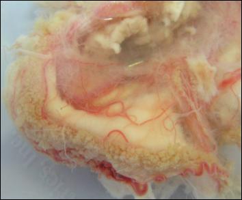
P. 1.
Vascular plexus of lateral
ventricle of a child 7,5 years. Macropreparations. zoom. 1,2х
1–
villiferous part; 2 – unvilliferous part;
3 – microarcads
![]()
![]()
![]()
![]()
![]()
![]()
![]()
![]()
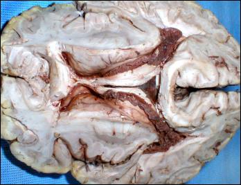
P. 2. General view
of the vascular plexus of lateral ventricle of the man 50 years. Macropreparations. zoom.
1:1
1– lateral ventricles; 2– villiferous part; 3 – unvilliferous part; 4 – brain tissue.
Therefore microvessels in
ontogenesis undergo changes which correlate with changes of the plexus itself
(that take place in it simultaneously with the development of the brain) and
reflect functional loads of the vascular plexus. The diameters of the
constituent parts of the microcirculatory channel of the vascular plexus of the
lateral ventricles of the brain increase (p<0.01) in mature age as compared with the
intrauterine period of the development 2.17 times.
Literature:
1. Anatomical characterization of human fetal brain development with
diffusion tensor magnetic resonance imaging / Hao Huang, Rong Xue, Jiangyang
Zhang [et al.] // The J. of Neuroscience. – 2009. – Vol. 1, № 29 (13). – P.
4263-4273.
2. Choroid plexus transplants in the treatment of brain diseases / Skinner
S.J., Geaney M.S., Rush R. [et al.] // Xenotransplantation. –
2006. – Vol. 13, № 4. – Р. 284-291.
3. Transplants of encapsulated rat choroid plexus cells exert
neuroprotection in a rodent model of Huntington's disease / Borlongan C.V.,
Thanos C.G., Skinner S.J. [et al.] // Cell Transplant. – 2008. – Vol. 16, № 10.
– P. 987-992.
4. Encapsulated living choroid plexus cells: potential long-term treatments
for central nervous system disease and trauma / Skinner SJ, Geaney MS, Lin H [et al.] // J. Neural. Eng. – 2009. – Vol.
6, № 6. – P. 65001.
5. Preterm brain ventricle reconstruction from 3-D ultrasound volumes / E.
Vansteenkiste, C. Metz, P. Govaert [et al.] // Processing of SPS-DARTS (the
third annual IEEE BENELUX/DPS Valley Signsl Processin Symposium). – 2007. – P.
167-172.
6.
Колесник В.В. Тривимірне
зображення та морфометричні параметри бічних шлуночків головного мозку
новонароджених / В.В.
Колесник, І.Ю. Олійник,
О.В. Корнійчук
// Вісн. проблем біол. і мед. – 2011. – Вип. 3, № 2 (88) – С. 88-92.