Shchepilina O.V., Begun P.I.
Saint Petersburg
Electrotechnical University «LETI»
Method research of the
system"thigh-bone graft-implant" in rehabilitation period after
osteosynthesis
A hip fracture is a heavy injury to the musculoskeletal system, this type
of injury is referred to hip fractures. Rehabilitation after hip replacement
surgery depends on many features and do not to say that there is one single
program. Modern problems of rehabilitation after hip fracture due to the fact
that is not governed by the maximum load, taking into account the recovery of
bone regenerate and is not considered a risk of vascular disorders. At the same time during postoperative period
after the hip fracture results from the fact that the thighbone traumatic injury
affects the locomotor system kinematic reactions in general, thus facilitating
associated disorders that do not directly result from the injury, yet worsening
the patient’s life.
Despite new implant designs, improved skills of
surgeons, new operation methods implemented, the results stop satisfying
patients as the full recovery period reaches half a year. This is because the
missing is the individual approach depending on the bone tissue condition, the
fracture location. The issue of the bone graft reconstruction at the subcapital
fracture location lacks attention.
However, information technologies development in
medicine, particularly in trauma surgery, orthopedics and biomechanics allows
achieving radically new rehabilitation technology level.
The object of the research is to develop
thighbone diagnostic technique after osteosynthesis with muscle activity and
elasticity module (E, MPa) taken into account at every bone graft remodeling
stage. The algorithm has been developed, the calculations have been carried out
and the analysis and the research have been undertaken for the “thighbone-bone
graft-implant” system stress and stain behavior at various rehabilitation
stages.
The following assumptions were considered while
building the conceptual model: 1) thighbone bone structure is idealized to
comprise two isotropic layers: cortical and spongy; 2) within the thighbone,
the fissure is located at the thighbone neck cross-section and it has uniform
isotropic structure, wherein its mechanical properties change at every
osteotylus reconstruction stage and those are localized within the zone that is
free of muscular efforts; 3) dynamic stress is applied to the thighbone center
by axes X, Y, Z (www.orthoload.com).
Figure 1 represents experimental data of the effective
load changes as a function of time (fig.1).
Fig.
1.
As initial data, the thighbone MRT is used
(fig.2) to build the object 3d models by means of Mimics, the computer modeling
environment. The figure 2 represents the thighbone (1 - spongy layer, 2 - cortical layer).
|
а |
b |
|
|
|
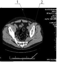
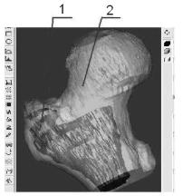
Fig.2
With those models imported into the Solid Works
software package, a solid thighbone geometric model was obtained with damages
at the area of the greater trochanter.
The considered is the bone recovery via
osteosynthesis, with two cannulated titanium screws (fig.3). The figure 3
represents the thighbone osteosynthesis (1 -
cortical layer, 2 - spongy layer;
3 - fixing screws, 4 - bone graft).
At every stage, the elasticity module is given
according to the diagram of the tab.1 that characterize the graft bone tissue
elasticity module change during the postoperative period.
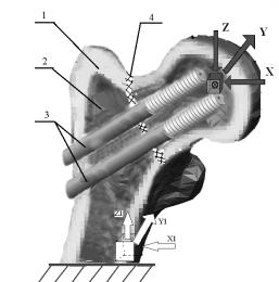
Fig.3.
Tab.1
|
№ |
Time after
operation, week |
Е, МPа |
|
1 |
before 3 |
0,0056 |
|
2 |
8-10 |
7,4 |
|
3 |
14-15 |
11,4 |
|
4 |
after 20 |
100 |
In terms of non-linear dynamic analysis, various
rehabilitation procedures, relating to the first two rehabilitation stages,
were considered. The obtained results are represented via fig.5. Fig. 4
represents dependences of deformations
appearing at the first stage with Eper=5.4kPa : 1 – allowed
deformation, 2 – deformation with the thigh aside, 3 – deformation for the
thigh up 30°.
Walking
is an important element in the complex process of rehabilitation
and positive effect on the work of many organs: cardiovascular
system, on the pulmonary system improves joint mobility, prevent muscle
degeneration.
Atrophy
of muscle tissue influences
the distribution of the load on
the system“thighbone-bone graft-implant”.We have made calculations based on muscle
atrophy which have a greater
impact on the distribution of the
load during walking.
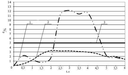
Fig.4.
The first stage of rehabilitation
In Fig.5.
represents dependences
deformation of the regenerate (E=6,62 MPa) from time double step when eighty
percent of muscle atrophy: 1) the quadriceps muscle of thigh; 2) muscle
antagonist synergist; position 3 on the chart of the femur is normal.
Fig.5
Method helps the surgeon in the choice of
technology operations and enables them to choose effective implant and method
of conducting rehabilitation program, knowing the risks.