DOES GLUTATHIONE IS A
SUFFICENT AGENT TO DEFENCE BRAIN AGAINST LIPID PEROXIDATION AFTER FEEDING RATS WITH
DIFFERENT FATS
Stępień Tomasz, Dziedzic Barbara, Świątek
Elżbieta, Walczewska Anna
Cell-to-Cell Communication Department,
92-215
Abstract
The aim
of our study was to investigate the effect of high-fat diet containing different
fats on lipid peroxidation, and glutathione (GSH) and GSH disulfide (GSSG) in
rat brains. Animals were fed the low fat and high fat diets prepared with the
same fat. Lard, as a source of saturated and monounsaturated fatty acids, sunflower
oil, as a source of linoleic acid (ω6 PUFA), and fish oil, as a source of
long-chain ω3 PUFAs. We determined concentrations of GSH, GSSG by Tietze
method and lipid peroxidation (LPO) products, malonodialdehyde plus 4-hydroxyalkenes
(MDA + 4-HDA), in brain homogenates. Six-week feeding with high amount of lard
and fish oil resulted in decrease in GSH and GSSG concentrations compare to the
corresponding low fat diets. High sunflower oil fed rats had equal level of GSH
compare to the low sunflower oil fed group. Oxidized-GSH had only tendency to
decrease in high sunflower oil fed rats. Fish oil in the low and high amount in
the diet was the most effective in lowering both GSH and GSSG concentrations. However,
the GSH-to-GSSG ratio in rats fed three different high fat diets did not differe
each other despite the differences in GSH and GSSG concentrations in these
groups. The GSH-to-GSSG ratio was enhanced in rats fed high lard compare to the
low lard diet. The level of LPO products
was a not compatible to the GSH-to-GSSG ratio in the brains. The both diets
rich in PUFAs, equally in the low and high amount in the diet, increased brain
LPO compare to the diet prepared with the same amount of lard. These results suggest
that lowering glutathione antioxidant efficiency, in part, may be responsible
for increased brain LPO in rats fed with fish oil but not in rats fed sunflower
oil diets. The underactivity of some other antioxidant enzymes or to low
concentration of other antioxidants may be a reason of a luck sufficient brain lipid
defense against an attack of oxygenic agents in these rats.
Introduction
A common Western diet characterizes a high amount of dietary fats. An amount and mostly used a type of fat in a diet depends on the world part, eating habits, social and family customs. A diet may be rich in saturated fatty acids (SFA), and monounsaturated fatty acids (MUFA) present in animal fats, like lard or beef tallow or in di- and tetra-unsaturated essential fatty acids, linoleic acid (LA; C18:2 ω6), and a-linolenic acid (ALA; C18:3 ω3) that cannot be formed de novo and should be ingested from a diet. These fatty acids are present manly in plant oils obtain e.g. from soybeans, olives or sunflowers. The essential fatty acids, and the complex lipids formed from them, are important constituents of biological membranes and contribute to maintain the structural and functional integrity of cells, and intracellular structures. Over the past decades grew up evidences of diverse beneficial effects of fish oil rich in omega-3 polyunsaturated fatty acids (ω3 PUFA) on heath (1). A dietary supplementation with eicosapentaenoic acid (EPA; C20:5 ω3) and docosahexaenoic acid (DHA; C22:6 ω3) is recommended for prevention of cardiovascular diseases (2) and cancer (3). However, unbalanced and high dietary fats may change metabolic pathways and affect many biological functions of tissues and organs. The type of dietary fat differentially affects the level of oxidative stress. High-saturated fat diet evokes hypercholestrolemy and enhances oxidative LDL modification (4). Omega-3 PUFAs in diet are incorporated in cell membranes, increasing the polyunsaturation of plasma membranes and their susceptibility for lipid peroxidation (5).
Reduced glutathione (GSH), a linear tripeptide of
L-glutamine, L-cysteine, and glycine is a relatively small ubiquitous molecule
in living cells (6). Its high electron-donating capacity (high negative redox
potential) combined with high intracellular concentration (millimolar levels) generate
great reducing power. (7) This characteristic underlies its potent antioxidant capacity
and defense neurons against oxidative stressors include ultraviolet and other
radiation (8), viral infections (9), environmental toxins, chemicals, and heavy
metals (7), inflammation, burns and septic shock (10). Deficiencies in brain glutathione
metabolism appear to be connected with several neurodegenerative diseases and
brain aging (11). In Alzheimer's disease a decrease in gluthatione level has
been reported and in Parkinson's disease the substantia nigra becomes greatly
depleted of GSH (12).
In the present study, we investigated the concentration
of malonodialdehyde plus 4-hydroxyalkenes
(MDA + 4-HDA), as an index of lipid peroxidation and the GSH status in brain
homogenates after six weeks rat feeding with three high-fat diets composed of the
different fats. Lard, as a source of SFA and MUFA, sunflower oil, as a source
of LA, (ω6 PUFA), and fish oil, as a source of long-chain ω3 PUFA.
Materials and Methods
Animals and diets
Male Wistar rats (150 –
Table 1. Composition of the experimental diets
|
|
Low-fat diet |
High-fat diet |
|
g/kg |
|
|
|
Casein |
194.4 |
228.6 |
|
Corn Starch 1 Sucrose |
364.4 291.5 |
252.6 201.1 |
Fat 2
|
52.5 |
203.4 |
|
Cellulose 3 |
48.6 |
57.1 |
|
AIN-93 Mineral Mix 4 |
34.0 |
40.0 |
|
AIN-93 Vitamin Mix 4 |
9.7 |
11.4 |
|
Choline bitartrate 5 |
1.9 |
2.3 |
|
L-Cystine 5 Energy density |
2.9 kcal/g 3.92 |
3.4 diet 4.62 |
1 – Stobimyl XMH 042 (Stockmeier Food, Germany); 2 – Lard, (Pamso S.A., Poland), Menhaden Fish Oil (Omega Protein, Inc. Hammond, LA, USA), Sunflower Oil, (Fat Processing Com. Inc, Warsaw; Poland); 3 – Arbocel â (J. Rettenmaier & Söhne Gmbh + Co Faserstoff-Werke, Rosenberg, Germany); 4 – Research Diets, Inc (NJ, USA); 5 – Sigma-Aldrich Sp z o.o
Table 2. Fatty acid composition of dietary fats
|
Total fatty acids |
Lard |
Sunflower oil |
Fish oil |
|
% by weight |
|||
|
Saturated |
42.6 |
9.2 |
29.0 |
|
Monounsaturated |
50.3 |
30.5 |
26.0 |
|
n-6 PUFA |
6.3 |
59.5 |
2.6 |
|
n-3 PUFA |
0.3 |
0.6 |
30.7 |
Glutathione assay
The brains were immediately removed from the sculls,
washed carefully in ice-cold PBS and homogenized in ice-cold 5% 5-sulphosalicilic
acid (1 ml/100 mg of tissue). Than homogenates were centrifuged at 10000xg for
10 min at 4°C, supernatants were collected and aliquots
were stored at -70°C. The Tietze`s GSH recycling method was used for
GSH determination (13). This method is based on an enzymatic recycling
procedure in which GSH is sequentially oxidized by
5,5’-dithiobis-2-nitrobenzoic acid (DTNB) and reduced by glutathione reductase
in the presence of NADPH. The rate of
formation of 2-nitro-5-thiobenzoic acid was spectrophotometrically measured at
412 nm and GSH was quantitated by reference to the standard curve. For total
GSH, the samples were added to the buffered solution (
Lipid peroxidation products
measurement
The
brains were homogenized in ice-cold
Chemicals and protein
determination
All chemicals used in the assays were supply by
Sigma-Aldrich Co. Protein content was measured using the
Statistical analysis
Data are shown as means ± SEM. ANOVA test followed by
Results
Lipid peroxidation
Rats fed both the low and high amount of lard revealed the lowest concentration of lipids peroxidation products (p<0.05; fig. 1). Any of the corresponding low fat and high fat fed groups did not differ significantly each other in brain lipid peroxidation. There were no significant differences in LPO level between rats fed with sunflower oil and fish oil in the low fat and high fat diet groups, as well.
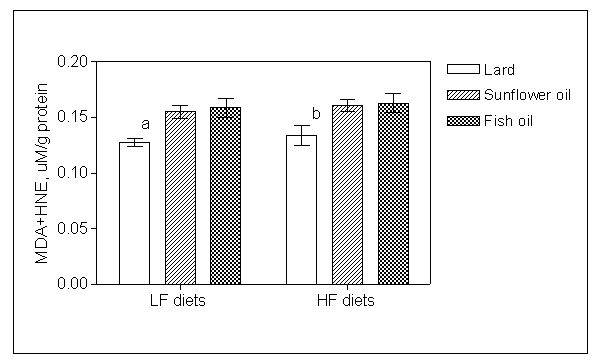
Fig.1. Effect of six weeks
feeding low fat (LF) and high fat (HF) diets composed of lard, sunflower oil
and fish oil on concentration of lipid peroxidation products in rat brains (n=12).
Values are means ± SEM. a p<0.05 vs. LF sunflower
and fish oil diets; b p<0.05 vs. HF sunflower and fish
oil diets.
Glutathione status
The comparison of the low fat and high fat diets showed differences within lard and fish oil fed rats. High lard and fish oil fed animals had a significantly lower concentration of GSH and GSSG than the corresponding low fat fed rats (p<0.05; fig.2). In turn, the high fat diet composed of sunflower oil did not alter the concentration of GSH compare to the corresponding low fat diet. A tendency to decrease in GSSG level by high sunflower oil was only noted. The highest concentration of GSH among the high fat diets fed rats was find in animals fed with high sunflower oil (p<0.01). Evaluating glutathione status in the brains of rat fed three different fats in low and high fat diet, GSH/GSSG ratio was increased significantly only in rats fed high lard compare to the corresponding low fat diet (p<0.05). The lowest brain GSH/GSSG ratio reviled rats fed the low lard diet (p<0.01).
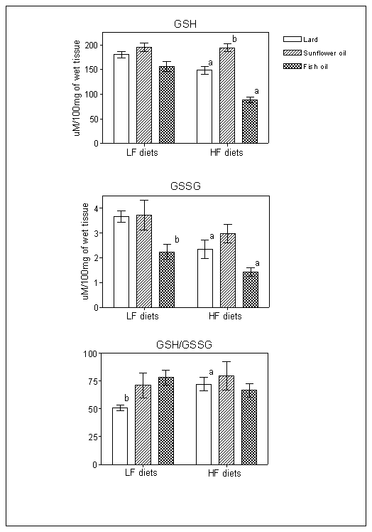
Fig. 2. Effect of six weeks feeding low fat (LF) and high fat (HF) diets composed of lard, sunflower oil and fish oil on glutathione (GSH) and oxidized glutathione (GSSG) concentrations in rat brain homogenates, and GSH/GSSG ratio (n=12). Values are means ± SEM.
a p<0.05 vs. the corresponding LF diet; b p<0.01 vs. the other LF or HF diets.
Discussion
Fatty acids are essential structural components of the central nervous system and play a role in its function, as well (17). However, grater amount of dietary PUFA causes their increased incorporation into bilayer membranes and enhances susceptibility to oxidation (5). The neuronal components are protected against its damage by enzymatic and non-enzymatic antioxidants. The enzymes include CuZn-superoxie dismutase and Mn-superoxide dismutase (SODs), GSH peroxidase and catalase, as well as a small antioxidant molecules, like glutathione, ascorbic acid, vitamin E, and a number of dietary flavonoids (18).
Glutathione is an extremely important antioxidant cell
protector. It directly quenches reactive hydroxyl free radicals, other oxygen-centered
free radicals, and radical centers on DNA and other biomolecules (2). Deficiency
in brain glutathione metabolism, which suggests an appearance of oxidative
stress in neurons, is connected with several neurodegenerative diseases (11).
Moreover, a shifting the GSH-to-GSSG ratio towards the oxidizing state
activates several signaling pathways including protein kinase B, calcineurin,
nuclear factor kappa-B, c-Jun and mitogen-activated protein kinase, may result
in changes of brain function, reducing cell proliferation and increasing
apoptosis (16). Gluthathione is also a cofactor of (1) multiple peroxidase enzymes that detoxify
peroxide products generated from oxygen radical attack on biological molecules;
(2) transhydrogenases that reduce oxidized centers on DNA, proteins, and other
biomolecules; and (3) glutathione S-transferases that conjugate GSH with
endogenous substances and diverse xenobiotics.
Our study showed that both ω3 and ω6 PUFAs independent on amount in the diets increased the concentration of the lipid peroxidation products in brain compare to the lard diet but the GSH-to-GSSG ratio in brains of rats fed three high fat diets was similar. However, fish oil fed rats, especially in high amount in the diet, reviled significantly lower both GSG and GSSG concentrations although the GSH-to-GSSG ratio in these rats was comparable to sunflower oil fed rats. It means that a GSH-to-GSSG ratio may be a not specific index of intracellular antioxidant capacity. The present study clearly demonstrates that, at least in dietary ω6 PUFA, the lowering of glutathione level was not responsible for increased LPO in brain. It arise a question which other antioxidant system may defense brain lipids against their oxidation in case of increased fatty acid polyunsaturation of their cell structure membranes. Based on our study we cannot answer on this question. However we speculate two possibilities. First, any of normal working intraneuronal antioxidant systems is not able sufficiently defense plasma membranes against an attack of free radicals and oxidative agents without an increase in other antioxidant concentration e.g. vitamins E and C. Second, one or more other intraneuronal antioxidant systems were affected by enhanced level of dietary PUFA and it was less productive in cell detoxification from an oxidative attack on lipids. An underactivity of GSH peroxidase, catalase and/or SODs that could increase in brain oxidative agents concentration should be consider.
References
- Ruxton CH,
- Weber HS, Selimi D, Huber G.
Prevention of cardiovascular diseases and highly concentrated n-3
polyunsaturated fatty acids (PUFAs). Herz. 2006,
- Gutt CN, Brinkmann L, Mehrabi A, Fonouni H, Muller-Stich BP, Vetter G, Stein JM, Schemmer P, Buchler MW. Dietary omega-3-polyunsaturated fatty acids prevent the development of metastases of colon carcinoma in rat liver. Eur. J. Nutr. 2007, Jun 25.
- Korpela R, Seppo L, Laakso J, Lilja J, Karjala K, Lahteenmaki T, Solatunturi E, Vapaatalo H, Tikkanen MJ. Dietary habits affect the susceptibility of low-density lipoprotein to oxidation. Eur. J. Clin. Nutr. 1999, 53(10):802-7.
- Yuan YV, Kitts DD. Dietary (n-3) fat and cholesterol alter tissue antioxidant enzymes and susceptibility to oxidation in SHR and WKY rats. J. Nutr. 2003, 133:679-688.
- Wu G, Fang Y-Z, Yang, Lupton JR, Turner ND. Glutathione metabolism and its implications for health. J. Nutr. 2004, 134:489-492.
- Kidd PM. Glutathione: systemic protection against oxidative and free radical damage. Altern. Med. Rev. 1997, 1:155-176.
- Cai J, Nelson KC, Wu M, et al. Oxidative damage and protection of the RPE. Progr. Retinal. Eye Res. 2000, 19:205-221.
- Look MP, Rockstroh JK, Rao GS, et al. Serum selenium, plasma glutathione (GSH) and erythrocyte glutathione peroxidase (GSH-Px)-levels in asymptomatic versus symptomatic human immunodeficiency virus-1 (HIV-1)-infection. Eur. J. Clin. Nutr. 1997, 51:266-272.
- Spies CD, Reinhart K, Witt I, et al. Influence of N-acetylcysteine on direct indicators of tissue oxygenation in septic shock patients: results from a prospective, randomized, double-blind study. Crit. Care Med. 1994, 22:1738-1746.
- Bains JS, Show CA. Neurodegenerative disorders in humans: the role for glutathione in oxidative stress-mediated neuronal death. Brain Res. Rev. 1997, 25:335-358.
- Tietze F. Enzymatic method for
quantitative determination of nanogram amounts of total and oxidized
glutathione: applications to mammalian blood and other tissues. Anal. Biochem. 1969, 27:
502-522.
- Sen CK. Cellular thiols and redox-regulated signal transduction. Curr. Top. Cell Regul. 2000, 36:1-30.
- Kitajka K, Sinclair AJ, Weisinger RS et al. Effects of dietary omega-3 polyunsaturated fatty acids on brain gene expression. PNAS 2004,101:10931-10936.
- Monks TJ, Ghersi-Egea J-F, Philbert M, Cooper AJL, Lock EA. The role of glutathione in neuroprotection and neurotoxicity. Toxicological Sci. 1999, 51:161-177.