The steps of treating a
patient with chronic traumatic arthritis of the temporomandibular joint.
Semenov К.А., Stepanova
S.V., Fesenko V.I., Ignatenko S.P.*
The State Institution
“Dnipropetrovsk Medical Academy of HM Ukraine”
The
Municipal Institution “Dniprodzerzhinska Dental Clinic ”DRC”*
Chronic arthritis - inflammation of the
temporomandibular joint (TMJ), flowingon the background of the destructive
changes in the bony structures of the temporomandibular joint [2,3,4,5].
Chronic arthritis is characterized by moderately
severe pain in the TMJ, often triggered by hypothermia and developed after a
lengthy speech load or during a meal (in case of solid food). The following is
also noted: stiffness in the joint in the morning and at the end of the day, stiffness
when chewing or talking. Limitation of mouth opening is observed, mainly in the
period of exacerbation. Radiographs reveal areas of narrowing or widening of
the joint space, the centers of osteoporosis and bone destruction of TMJ joint
elements [4].
Abnormalities in the dentition are the starting
point in the development of TMJ arthritis. Painful TMJ
dysfunction is the main reason of chronic arthritis. A.Mirza explains the
pathogenesis of pain dysfunction as follows [4]. In cases when during clamping dental
arches individual teeth or areas of the occlusal surface come into contact
before the others (premature contacts) barriers occur for clamping other teeth
[1,5]. This can lead to a shift of the lower jaw in a forced position when
multiple contacts are achieved at the cost of violation of coordinated
functions of the masticatory muscles and changing the position of the articular
heads. This leads to disruption of normal bilateral function of the masticatory
muscles and temporomandibular joint elements and clinically causes painful
dysfunction. Reflexively the function of masticatory muscles changes, the
process of chewing rebuilds, which leads to microtraumas of joint elements and
disturbance of their trophicity.
Clinical studies conducted by A. Mirza proved
that with the ongoing changes in the relative position of the articular head,
holes and disk the functional load is redistributed to the various sections of
the joints. In areas where the load is increased, there is compression of the
soft tissues, in other areas with stretches - distraction. The result is the
formation of cracks, perforation, ruptures of the disc and articular cartilage,
articular ligament sprains, dislocation of the diosk, trophic changes in the
joint.
Consider a clinical example: the patient K. born
1989 complained on difficulties in opening the mouth (mouth opening distance
was 1.5 cm). Upon visual inspection a slight asymmetry of the face on the right
side was spotted. The cause was the hypertrophy of the masseter muscle, which is
a characteristic of one-side chewing.
It was recommended for Patient K to do the
panoramic radiography. According to the panoramic radiography and examination
of dentition the following was found: changes in the shape and structure of the
head of the TMJ on the left, dystopia of teeth 18 and 28, lack of space for
impacted teeth 48 and 38, malocclusion in the frontal area due to reduced lower
jaw. The bite was deep and prognathic (second class first subclass
classification Engle). Teeth examination revealed: photopolymer restoration of
teeth 17, 16, 26, 27, 36, 35, 45, 46 and 46, and endodontic treatment of a
tooth 46. Established that a violation of occlusal relationship of the teeth of
the upper and lower jaw, the violation of a uniform distribution of masticatory
forces in the distal parts of the dentition (Fig. 1).
Based on complaints, and clinical and
radiographic examination the arthritis on the left TMJ was diagnosed.
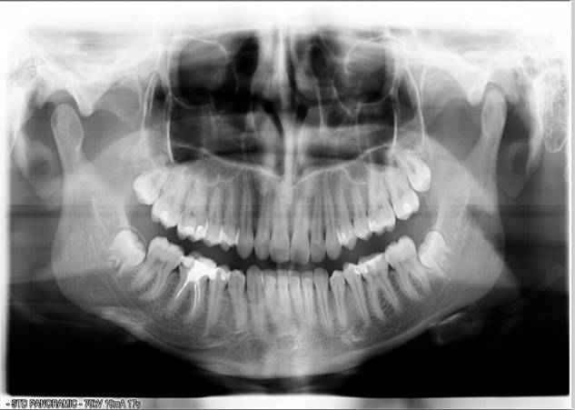
Figure 1
Panoramic radiography of patient K prior to treatment in 2008
Examination
and treatment of patient K. went on for two years. Therapeutic measures have
been divided into five stages, which include: medical therapy, surgical
sanitation - removal of dystopic teeth, orthodontic treatment, therapeutic
reorganization and orthopedic stabilization of the bite height. In the early
stages of treatment a medical therapy was held in order to relieve inflammation
and restore the function of the TMJ. In the scheme we developed the following
products were included:
1. Restructa
pro injectione C - 1 shot (i.m.), after 2 days, 2 ml syringe. 5 weeks
2. Dona - 1 amp. + Supplied
solvent, i.m., 3 times a week for 4 weeks
3.
Hondroksid (gel) - Lubricate the skin in the region of the joints 2 times a day.
4. Persen - 1 tab. 2 times a day.
Two month
drug therapy restored the full opening of the mouth and eased movements in the
left TMJ.
In the
second phase of treatment, patient K was encouraged to get removed dystopic teeth 18 and 28. After surgery and analysis of diagnostic
models one year orthodontic treatment was recommended.
In order to stabilize the height of occlusion after orthodontic treatment the following was recommended: therapeutic rehabilitation in the dentition,
orthodontic treatment, which consisted of a counter-crowns on teeth 16 and 46,
26 and 36, the removal of impacted teeth 48 and 38 (Fig. 2).
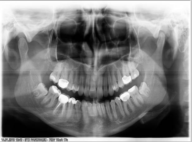
Figure 2 Panoramic radiography of patient K after orthodontic and orthopedic
treatment in 2010
At the moment, the patient K feels well,
no complaints about TMJ. In case of future painful
phenomena in the TMJ patient K was advised to use
the standard glenoid kappa for a month for 3 hours a day.
Conclusions
1. There
is no universal
method or ideal scheme applied to treat the diseases of the temporomandibular
joint yet. An integrated approach to pathology of the temporomandibular joint
that takes into account all the factors contributing
to the functional disruption of the joint allows to stabilize the development
of this disease and relieve the patient's condition during periods of exacerbation.
2. Accurate diagnosis taking into account
the etiological factors in the pathology of the temporomandibular joint allows
to select the most efficient method of treatment and contributes to long-term
remission of the disease.
3. Detection of infringements and the
normalization of the occlusal relationship of the teeth in the dentition is a
major challenge in the treatment of pathologies of the temporomandibular joint.
References
1. Гросс
М.Д. Нормализация окклюзии/
М.Д., Гросс, Дж.Д. Мэтьюс - М.: Медицина. – 1986. – 286с.
2. Ральф Е.,
Мак-Дональд, Дейвида Р. Эйвери. Стоматология детей и подростков /Е.Ральф,
Мак-Дональд, Дейвида Р. Эйвери. - М.: Медицинское стоматологическое агентство.-
2003.- 766 с.
3. Робустова
Т.Г. Хирургическая стоматология /Т.Г.Робустова - М.: Медицина. – 1996. - 688с.
4. Тимофеев А.А. Руководство по
челюстно-лицевой хирургии и хирургической стоматологи/ А.А.Тимофеев -
К.: ООО “Червона Рута-Турс”. - 2004. - 1062с.: ил.
5. Хватова В.А.Клиническая
гнатология/ В.А. Хватова - М: Медицина.- 2005. – 312с.