1Limanskaya L.A., 1Pakhomova
E.V., 1Trusova V.M., 1Gorbenko G.P.,
2Deligeorgiev T., 2Vasilev
A., 2Kaloianova S., 2Lesev N.
1V.N. Karazin
Kharkiv National University, 4 Svobody Sq., Kharkiv, 61077, Ukraine
2Department of Applied Organic Chemistry, Faculty of
Chemistry, University of Sofia, Sofia, 1164, Bulgaria
DELIVERY
OF NEW POTENTIAL ANTITUMOR DRUG BY LIPOSOMAL NANOSYSTEMS
Europium
chelate (EC) (here referred to as V10) belongs to a new class of potential
antitumor drugs with high cytotoxic activity. Lanthanide complexes are of
particular interest for biomedical investigations and diagnostics, since their
spectral characteristics are optimal for decrease of light scattering in
biological patterns and fluorescence background contribution. However, the application
of such drugs in a free form is limited by their high toxicity and metabolic
instability. One efficient way to increase drug efficiency is based on using
different drug delivery systems such as dendrimers, nanotubes, nanoshells,
quantum dots, liposomes, etc. Highly adaptable liposome-based nanocarriers
currently attract increasing attention, because of their indisputable advantages,
viz. complete biodegradability,
ability to carry both hydrophilic and lipophilic payloads and protect them from
chemical degradation and transformation, increased therapeutic index of drug,
flexibility in coupling with targeting and imaging ligands, improved
pharmacodynamic profiles compared to free drugs, etc.
The aim of current research
was: 1) identifying the probes whose fluorescence is quenched by EC; 2)
evaluating the most probable EC localization in liposomal membranes consisting
of phosphatidylcholine (PC) by comparing the quenching efficiencies for probes
differing in their location across the lipid bilayer. To achieve this goal, we evaluate
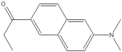
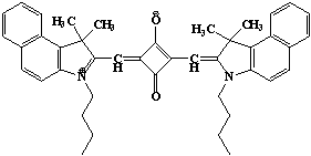
V10 as a quencher for the fluorescent probes Prodan, 4-dimethylaminochalcone (DMC), Laurdan, 3-methoxybenzanthrone
(MBA) and SQ-1, residing at different depths in the liposomal membranes.
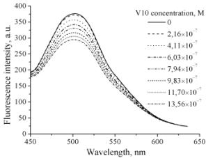
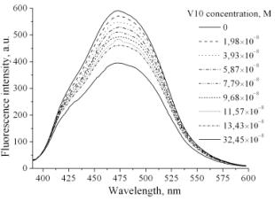
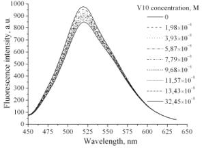
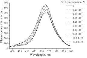
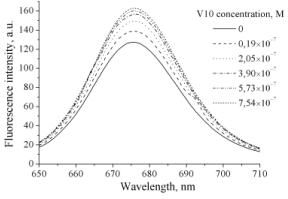
At the
first step of the study fluorescence spectra of DMC, Laurdan, MBА, Prodan and SQ-1 were recorded in the suspension of
PC liposomes in the presence of increasing concentrations of V10 (Fig.1).
(A)
(B)
(C) (D)
(E) (F)
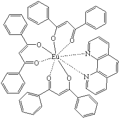
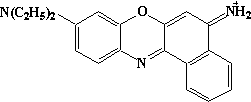
Fig 1.
Structural formula of europium chelate (A) and fluorescence spectra of DMC (B),
Laurdan (C), MBА (D), Prodan (E) and
SQ-1 (F) in suspension of PC liposomes in the presence of europium chelate V10.
As seen
from Fig. 1, addition of europium chelate was followed by the decrease in fluorescence
intensity of DMC, MBA, Laurdan and Prodan, only for squaraine probe SQ-1 an
opposite effect takes place. These data were interpreted in terms of EC ability
to serve as a quencher of DMC, Laurdan, MBА and Prodan
fluorescence.
Fluorescence quenching
has been widely studied both as a fundamental phenomenon, and as a source of
information about biochemical systems. Both static and dynamic quenching
requires molecular contact between the fluorophore and quencher. Collisional
quenching of fluorescence is described by the Stern-Volmer equation:
![]() ,
,
where F0 and
F are the fluorescence intensities in the absence and presence of a quencher,
respectively; km stands
for the bimolecular quenching constant; τ0
is the lifetime of the fluorophore in the absence of a quencher, and [Q] is the concentration of a quencher.
By analyzing the obtained spectra according to the Stern-Volmer equation, we
received Stern-Volmer plots (Fig. 2). Likewise, bimolecular quenching
constants which reflect the efficiency of the quenching
or the accessibility of the fluorophores to the quencher have been
evaluated (Table 1).
Table 1. Quenching parameters of fluorescent probes
|
Fluorescent probe |
Bimolecular
quenching constant, M-1×sec-1 |
|
DMC |
(6.1±1.7)×109 |
|
Prodan |
(2.7±0.8)×109 |
|
Laurdan |
(6.8±1.9)×1010 |
|
MBA |
(3.8±1.1)×1010 |
The quenching
efficiency of the probes was found to decrease in the order
Laurdan>MBA>DMC>Prodan. Since Laurdan adopts the deepest location in
liposomal membranes, embracing the glycerol backbone and initial acyl chain
carbons, it can be assumed that europium chelate under study, being nonpolar in
nature, partially penetrates in the hydrophobic region of the lipid bilayer.
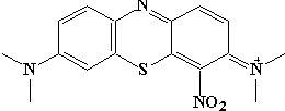
(A)
(B)
(С) (D)
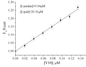
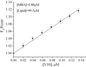
Fig 2. Structural formulas of
the probes employed: DMC (A), MBA (B), Prodan (C), SQ-1 (D), and typical
Stern-Volmer plots for fluorescence quenching by europium chelate V10 in suspension
of PC liposomes.
This work was supported in part by the grant #4534
from the Science and Technology Center in Ukraine and Fundamental Research
State Fund (project number F.28.4/007).
References
1. Lakowicz J.R.
Principles of Fluorescent Spectroscopy, third ed. Plenum Press, New York. 2006.
2. Zhang X., Lei X., Dai H. Synthesis and
characterization of light lanthanide complexes with 5-aminosalicylic acid //
Synth. React. Inorg. Met.-Org. Chem. 2004. V. 34(6). P. 1123-1134.