Медицина/6. Экпериментальная и
клиническая фармакология
Candidate of Pharm. Sc. Butko Y. O.
National University of Pharmacy, Ukraine
Effect of lotion with
glucocorticosteroid and ceramides on skin morphostructure in allergic contact dermatitis
High
level of dermatitis diseases in Ukrainian population, increasing the number of
severe clinical forms, and shortening the term of remission and low indicator
of recovery make a problem of dermatitis therapy, which is the one of topical
problems in sophisticated dermatology [2, 5].
Currently, the treatment of dermatoses
paid much attention to the state of proliferation of skin and restore its barrier function, because sound is the use of tools that stimulate proliferation and restore skin structure, including ceramides [4].
The aim of this work was morphological study of
rats skin state in treating by lotion "Mometasone with ceramides" in the course of allergic
contact dermatitis (ACD).
Materials and methods. The
object of study was animals skin after treating with lotion “Mometasone with
Ceramides”. 18 rats were used in study, which were divided on: 1 group – intact,
2 group – positive control (control pathology), 3 group – rats, which was
treated with lotion “Mometasone with Ceramides”. To develop AСВ animals were coated 5% alcoholic solution of
2,4-dinitro-chlorobenzene (DNCB) by the method of P. Zalkan [1]. Lotions were
applied a thin coat once a day. After 7th treating day animals were
removed from study and all skin material were fixed in 10% formalin solution
for morphological studying. Than skin examples were swamped in celloidin-paraffin
and sections were painted in hematoxylin and eosin (1) [3]. We performed review micropreparations on a microscope
Micros400 (Austria). Photomicrography
microscopic images was performed a digital camera Nikon Col Pix 4500. Pictures were
processed on a computer Pentium 2,4 GHz using Nikon View 5.
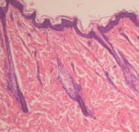
Results
and discussion. As we can see in studies of intact rats, epidermis developed
well, consists of 3-5 rows of cells. We differentiated in it: one row of basal
cells, 1-3 rows of thorny cells, one or rarely two rows of granular cells and
the stratum corneum, which consists of fluffy corneal plates. Derma is wide
with compact bundles of collagen fibers with a few thin-walled blood vessels. Cell
saturation dermis is mild (lymphoid cells, fibroblasts). Epidermal-dermal
border is clear. Hair follicles are multiple, cut as extended and cross section
(pic.1а).
|
|
|
|
|
а |
b |
c |
|
|
|
|
|
d |
f |
g |
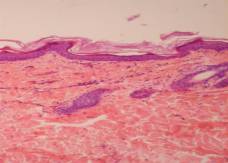
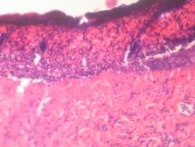
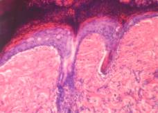
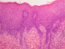
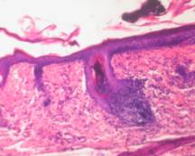
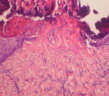
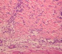
Рiс.
1 – intact rats skin (а). Positive control group rats skin: b – epidermis and
papillary dermis layer necrotized, demarcation shaft on the border with mesh
layer of the dermis; c – regenerated epidermis thickened under the crust, there
are visible signs are not sharply distinct spongiosis, fiber dermis are
swollen. d – hypertrophy of the epidermis, acanthosis, inflammatory reaction in
the dermis; f – inflammatory infiltrate around the hair follicle with the
destruction of the wall; g – narrowing of the capillary lumen, endothelial
proliferation, inflammatory reaction in the subcutaneous tissue.
Hematoxylin-eosin. х 100.
On 7th
day after applying crucial dose DNCB in all control pathology rats we found a
spreading skin destructions. Epidermis is deformed on this parts, often it
looks like amorphous mass, which densely infiltrated a cellular detritus, macrophages.
Collagen fibers are homogenized in papillary layer of the dermis, strongly
eosinophilic, epithelial cells of hair follicles and sheaths are in the
necrosis. There is visible demarcation shaft some places among the crust and
saved part of the dermis (pic. 1b). Often
crust extends far beyond the damage and covers (peels tightly or partially)
thickened epithelial layer, in which we could see a mild distinct spongiosis
(intercellular edema in the thorny layer), vacuolar degeneration of
epidermocytes. The epithelium of hair follicles proliferates. Collagen matrix
is swollen in the dermis and moderate polymorphonuclear infiltrates are visible
around some hair follicles and blood vessels (pic. 1c). Probably it was the
regeneration of damaged during primary-contact reactions to allergen skin in
these areas at the time of the experiment.
Often epithelial layer hypertrophied on the skin, free from peel, we can incomplete keratinization, acanthosis, more expressive spongiosis. We observed in the dermis infiltrates of histiocytes, lymphocytes, macrophages, plasma cells (pic. 1d). Inflammatory cell infiltration was observed often and around hair follicles and combined with their sebaceous glands (epithelial cells of them are necrotic) and in subcutaneous tissue. We traced wall thickening by hypertrophy and hyperplasia of endothelial cells and pericapillary in mesh dermis capillaries, capillary lumen is diminished (pic. 1f,g). The morphological picture is characteristic ACD.
During
the treatment lotion improved skin condition in 83.33% of the rats. Morphological
characteristics AKD were not in 33.3% of these animals (pic. 2а). On other 50%
of the rats was observed (mostly) small, limited both in depth and across the
injury site, covered the crust that is quite easy to flake off. We traced
obvious signs accelerating regeneration of the epithelium in these areas (expressive
as boundary epithelization and from the epithelium of saved hair follicles).
There is no or there is reduced inflammatory cell reaction in the dermis under
the, the condition of the walls and the size of the lumen were normal of most
blood capillaries (pic. 2b).
|
|
|
|
|
a |
b |
c |
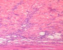
Рiс
2. Rats skin which were treated with lotion “Mometasone with Ceramides”: а – no
signs of allergic dermatitis; b – limited area of damage, no
inflammatory cell reaction in the dermis, the condition of capillaries is
normal; c – in the dermis condition of capillaries is close to normal,
inflammatory reaction precapillary and subcutaneous tissue is decreased.
Hematoxylin-eosin. х 100.
Spreading
peel outside reduced markedly damage zones. Crust is sequestered often, and
large areas of the skin surface are clean. The thickness of the epithelial
layer is clearly reduced relative to control pathology, signs of acanthosis
were not found. Sometimes there was a temperate spongiosis and vacuolar
dystrophy of epidermocytes. In the dermis condition of collagen matrix,
arteriovenula bridge, the hair follicles are close to normal, inflammatory
reaction is absent or greatly reduced (pic. 2c). Only one rat (16,7%), which
was treated with lotion “Mometasone with Ceramides”, skin condition was a
little different from that of the control rats.
Conclusion. Thus, we can concluded on the
basis of these studies, that rats have clear allergic skin inflammation after
applications with DNCB. Application of lotion “Mometasone with Ceramides” reduces
the intensity of ACD (50% rats did not have destructive epidermis changes, the
zone of damage is limited, reduced exudative manifestations and inflammatory
reaction in the dermis, signs of accelerating epithelial; 33,3% rats had normal
skin). Thus, the introduction of ceramides
to the cream of
mometasone furoate helps normalize skin structure that makes it worthwhile to use in the treatment of allergic dermatitis.
References
1. Бунятян Н.Д.
Эффективность 5 % альтановой мази при контактном дерматите у крыс / Н.Д.
Бунятян, В.В. Березнякова, Т.Ю. Глазкова // Вест. ВГУ. Серия: Химия. Биология.
Фармация. – 2004. – № 1. – С. 160-162.
2. Волкославська
В.М. Стан захворюваності на дерматози в Україні через 20 років після аварії на
ЧАЕС / В.М. Волкославська, О.Л. Гутнєв // Укр. журн. дерматол., венерол.,
косметол.: Наук.-практ. видання. – 2010. – № 3. – С. 153.
3. Меркулов Г.А.
Курс патологогистологической техники. – М.: Медицина, Ленингр. отд-ние, 1969. –
424 с.
4. Ceramides
and barrier function in healthy skin / J.Mutanu Jungersted, L.I.Hellgren, J.K.
Høgh [et al.] // Acta Derm Venereol. 2010. – Vol.4, № 90. – P.350-353.
5. Sehgal V.N.
Atopic dermatitis: current options and treatment plan / V.N. Sehgal, G.
Srivastava S. Dogra // Skinmed. – 2010. – Vol.8, № 6. – Р. 335-344.