USE OF ELECTROPHORESIS IN DIAGNOSIS OF CATTLE HUMORAL IMMUNITY
Małgorzata
Szewczuk1, Danuta Czernomysy-Furowicz2, Ewa
Czerniawska-Piątkowska1
Departament of Ruminant
Science1 and Departament of Immunology and Microbiology2,
Agriculture
ABSTRAKT: The
aim of the study was to determine the differences in electrophoretic assays and
concentration of total protein and its fractions in blood serum of
Black-and-White calves with varied addition of Holstein-Friesian genes. The
material comprised 148 samples of serum obtained from whole, non-homogenised
blood of calves, collected before 5 days of age and at age 3 months.
In
the calves that showed clinical symptoms of diarrhoea, a clear increase in
alpha2 fraction was observed and a slight increase in alpha1.
The concentration of albumin, and thus total protein, was reduced. Increased
globulin fractions accompanied by decreased albumin concentration demonstrated
a bacterial infection.
In
a pneumonia-affected calf's serum, a significant increase in alpha1 and alpha2
fractions was found with a slight increase in beta2 fraction. The
increase of both alpha fractions and beta2 fraction is observed with
enhanced cellular non-specific response to the disease process. Due to a slight
increase in the gamma fraction increase, it can be concluded that M-class
immunoglobulins have been synthesised.
An
electrophoretic assay, which allows measuring concentrations of individual
fractions and albumin-globulin ratio, represents a powerful that can be used in
evaluation of immunity and health of livestock animals.
Keywords: calves,
electrophoresis, health, acute phase proteins
INTRODUCTION
Electrophoresis, which allows us to determine the
concentration of each faction and albumin to globulin ratio, is an important
assay that can be used in assessment of immunity and health of livestock
animals.
Very often, a lack of clinical symptoms in sick
animals may lead to an epizootic, which is particularly dangerous within first
months of life of calves managed in large herds. Electrophoresis of blood serum
and the analysis of proteinograms allows us to picture the health condition of
the animals, also in terms of therapeutic success. The proteins form particular
fractions. As a result of inflammation, we observe an increase in the
concentration of acute phase response proteins and, consequently, increased
concentration of the fractions that contain acute phase proteins (APP). The
role of APP in the acute phase response consist in quenching the inflammation
process, eliminating the damage factor, healing the damaged tissues and organs,
and thus restoring the organism homoeostasis (Kostro et al., 2002). Increased
concentration of APP is reflected in the plasma electrophoretic assay in the
form of increased concentration of, among others, alpha1 and alpha2
globulin fractions (Dembińska et al., 2002). Under a disease condition,
the concentrations of haptoglobulin (HP), serum amyloid A protein (SAA),
fibrinogen (Fb), alpha-1-acid glycoprotein (AAG), antitrypsin (AT), c-reactive
protein (CRP), and alpha2 macroglobulin (MG) increases
(Czokała-Plichta, 2002), whereas the concentration of albumin (Alb) and
transferrin (Tf) decrease (Czokało-Plichta, 2002, McNair et al., 1998,
Kent, 1992).
Determination of the concentration of fractions and
APP in the animal blood serum allows monitoring their health, which enables
early detection of inflammations and post-clinical infections – the conditions
that reduce weight gains, deteriorate the animal performance, and lead to an
economic loss (Kostro et al., 1996; Heegaard et al., 1998;
Włodarczyk-Szydłowska et al., 2000). Moreover, altered concentrations
of these proteins in the blood serum of beef cattle represent an indication of
faulty conditions in terms of welfare (Kostro, 2002).
MATERIAL
AND METHODS
The studies took place during 2000-
The material comprised 148 samples of blood serum
collected from calves. The serum was obtained from whole blood,
non-homogenised, collected from the external jugular vein on the 5th
day after birth and at age 3 months. Total serum protein concentration was
measured with burette method. The serum electrophoresis was performed in HR7
hydrogel using a Cormay Diagnostics chamber, whereas the concentrations of
fractions were measured in a DS-3 densitometer (Cormay). The values of total
protein content (in g/l) were used to convert the percentage of each fraction
into their concentrations in g/l.
During the first 3 month of life, the calves were also
examined for health. The recorded disease units were divided into three groups:
airways diseases (pneumonia, bronchopneumonia), alimentary tract diseases
(diarrhoeas of various origin), dermal diseases (mycoses).
RESULTS
AND DISCUSSION
The following fractions were found in the studied
calves: 1 albumin and 7 globulin fractions, i.e. alpha1, alpha2,
alpha3, beta1, beta2, gamma1, and
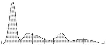
gamma2. Albumin was the dominant fraction for healthy calves (Figure
1). No increased concentrations of globulin fractions were found, neither any
additional peaks that would demonstrate enhanced synthesis of APP.
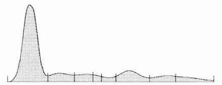
albumina α1 α2 α3 β1 β2 γ1 γ2
Ryc. 1.
Proteinogram surowicy zdrowego cielęcia
Figure 1. Proteinogram of a healthy calf serum
The
resulting proteinograms for ill animals showed changes in the fraction levels
and additional proteins:
Changes at fraction level
The proteinograms of calves that
underwent homoeostasis disturbances look entirely different (Figure 2). Calves
with clinical diarrhoea symptoms showed an explicit increase in alpha2 and a
slight increase in alpha2 fraction accompanied by reduced concentration of
albumin and, consequently, total protein. In various diseases which run with
increased permeability of capillaries, albumin tends to escape into the
extravascular space, which is accompanied by a drop in the protein serum
concentration. Increased globulin fractions accompanied by reduced albumin
level may indicate a bacterial infection, which has also been confirmed on
calves by Schneider (2003).
B A
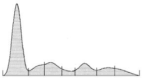
albumina
α1 α2 α3 β1
β2 γ1 γ2 albumina α1
α2 α3 β1
β2 γ1 γ2
Ryc. 2.
Proteinogramy surowicy cieląt z objawami biegunki
Figure 2. Serum proteinogram of a calf affected
with diarrhoea
Increased concentration of both
gamma-globulin fractions is typical for polyclonal hypergammaglobulinaemia
(Figure 3). Changes of this kind occur during growth processes that are
characterised by immunological complexes formed by homogeneous immunoglobulins
with molecules of other immunoglobulins or with any other serum proteins. The
fraction gamma2 revealed also an additional peak, characteristic for monoclonal
hypergammaglobulinaemia. Enhanced synthesis of antibodies by a single B
lymphocyte clone can be observed with viral infections. Bacterial infections,
in response to which the synthesis of alpha fractions increases, represent a
binding agent for viral infections. Therefore, it can be assumed that the calf
which revealed clinical symptoms of bronchopneumonia had undergone through
viral infection in the first place, followed by a bacterial infection.
![]()
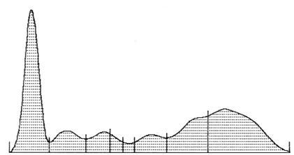
albumina α1 α2 α3 β1 β2 γ1 γ2
Ryc. 3.
Proteinogram surowicy cielęcia z objawami bronchopneumonii
Figure 3. Serum proteinogram of a calf with
symptoms of bronchopneumonia
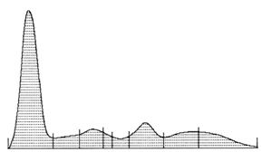
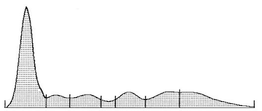
Figure 4 presents a proteinogram of
serum collected from a pneumonia-affected calf. The blood was drawn on the day
following the moment when the clinical symptoms had been observed. The serum of
the calf contained a considerably elevated levels of the alpha1 and
alpha2 fractions. The alpha1 consists of alpha1-antitrypsin
and alpha1-acid glycoprotein, while alpha2 contains
haptoglobulin and alpha2-macroglobulin. A slight increase in beta2
fraction, which contain beta-lipoprotein and C3 complement factor, was also
observed (Bigoszewski et al., 2001; Dembińska, 2002; Kostro et al., 1996;
2001; 2002). An increase in both alpha fractions and beta2 fraction
accompanies an increase in non-specific cell response to the disease. Due to a
small increase in the gamma fraction, which is formed from immunoglobulins, it
may be concluded that synthesis of M-class immunoglobulins has occurred.
albumina α1 α2 α3 β1 β2 γ1 γ2
Ryc. 4. Proteinogram surowicy cielęcia w
początkowym okresie pneumonii
Figure 4. Serum proteinogram of a calf in the
initial stage of pneumonia
Presence of additional proteins
As a result of infection, the
synthesis of immune proteins, which stimulate cellular immunity, accelerates.
This enhanced synthesis is manifested in the electrophoretic assay not only
through an increased concentration of the fraction, but with additional peaks
as well.
![]()
albumina α1 α2 β1 β2 γ1 γ2
Ryc. 5.
Proteinogram surowicy cielęcia z widocznym dodatkowym białkiem
Figure 5. Serum proteinogram of a calf with an
additional protein
During the first 24 hours of pneumonia,
serum revealed an increased concentration of c-reactive protein. This protein
binds to pneumococcal C polysaccharide, and also activates the classic
complement pathway. Serum electrophoretic assay of these calves has revealed an
additional peak between beta and gamma fractions (Figure 5). An increase in the
protein concentration sometimes precedes clinical symptoms (Bigoszewski et al.,
2001; Dembińska, 2002; Kostro et al., 2001; 2002). It is also possible
that the fraction also contains A immunoglobulins, whose increase is observed
during the first days of infection.
Serum amyloid A protein (SAA)
represents one of the first proteins of the acute phase (Bigoszewski et al.,
2001; Dembińska, 2002; Kostro et al., 1996; 2001; 2002). An intensive
increase in the concentration of this protein is observed within 20 hours after
the traumatic agent activates. In a few day-old calf (Figure 6), which had not
revealed any clinical symptoms, SAA could have originated from the dam, in
which the concentration of this protein had increased during gestation.
![]()
albumina α1 α2 β1 β2 γ1 γ2
Ryc. 6. Proteinogram surowicy 3 dniowego cielęcia
z widoczną zmianą frakcji albuminowej wywołane pojawieniem
się białka SAA
Figure 6. Serum proteinogram of a tree day-old calf with a visible
change in the albumin fraction resulting from the occurrence SAA
Local or general immunity suppression
caused by endogenous factors (cancerous and autoimmunological diseases) or
exogenous factors (infections, stresses, contamination of environment) leads to
development of diseases caused by opportunistic microorganisms. Whether we
succeed to prevent these diseases heavily depends on the proper health
monitoring of the animals. One of the way to evaluate homoeostasis of the
animals is to control the behaviour of the acute phase proteins (APP). Surveying
the serum APP concentration within the monitoring of livestock animals health
enables early detection of homoeostasis disturbances and a quick decision can
be reached in order to its restitution (Bigoszewski et al., 2001; Kostro et
al., 2001 after Hedstron et al.; Stefaniak, 2000). According to Kostro et al.
2001, measuring APP is particularly useful for identification of inflammations
developing with non-symptomatic infections, which are difficult to diagnose.
Nevertheless, proteinograms provide visualisation of both fractions
concentration changes and APPs. This method may be applied before we assay
specific APPs.
Plotting serum proteinograms for the animals, and thus monitoring of
their health, allows us to detect disease conditions before clinical symptoms
arrive, a quick veterinary treatment, and to avoid an economic loss. Monitoring
the immunity of the animals by electrophoresis is of a great practical value
and can be readily carried out. Blood may be collected along with official
samplings, which does not disturb the farm operations; additionally, the ease
of performance and readily available results represent another argument
supporting a wider application of proteinograms in cattle disease diagnostics.
REFERENCES
Bigoszewski M., Rychlik A., Depta A. (2001): Białka
ostrej fazy u zwierząt. Med. Wet., 57(3): 151-155.
Czokało-Plichta M. (2002): Białka osocza i
hemostaza W: Patofizjologia, Maśliński S., Ryżewski J.,
Wydawnictwo Lekarskie PZWL, Warszawa.
Dembińska – Kieć A., Drożorż R. (2002):
Białka osocza W: Diagnostyka laboratoryjna z elementami biochemii
klinicznej, Dembińska-Kieć A., Nasklaski J. W, Urban & Partner,
Wrocław.
Heegaard P.M., Klausen J., Nielsen
J.P., Gonzales-Ramon N., Pineiro M., Lampreave F., Alava M.A. (1998): The
porcine acute phase response to infection with Actinobacillus pneumoniae.
Haptoglobin, C-reactive protein major acute phase protein and serum amyloid A
protein are sensitive indicators of infection. Comp. Biochem. Physiol., 119B:
365-373.
Kent J. (1992): Acute phase
proteins :their use in veterinary diagnosis. Br. Vet. J.,
148: 279-282.
Kostro K, Sobieska M, Wiktorowicz K, Wołoszyn S. (1996):
Białka ostrej fazy u zwierząt – występowanie i charakterystyka,
Med. Wet., 52 (3): 152–153.
Kostro K., Gliński Z., Wójcicka- Lorenowicz
K., Krakowski L. (2001): Białka ostrej fazy jako markery chorób u
zwierząt. Med. Wet., 57(8): 539-542.
Kostro K., Wójcicka-Lorenowicz K., Gliński
Z., Krakowski L., Wrona Z. (2002): Białka ostrej fazy jako ligandy
komórek układu immunologicznego. Med. Wet., 58(12): 929-933.
Mcnair J., Elliott C., Bryson
D.G., Mackie D.P. 1998: Bovine serum transferrin concentration during acute
infection with Haemophilus somnus. Vet.J., 155: 251-255.
Schneider G. (2003): Kształtowanie się wybranych
elementów odporności humoralnej u cieląt do 12 miesiąca
życia pochodzących z różnych gospodarstw. Praca
magisterska. AR w Szczecinie.
Stefaniak T. (2000): Białka ostrej fazy w
diagnostyce u bydła. Zesz. Nauk. AR Wrocław, 390: 49-59.
Włodarczyk-Szydłowska
A., Chełmońska- Soyta A., Nowacki W. (2000): Białka ostrej fazy
u koni. Zesz. Nauk. AR Wrocław, 390: 69-74.
Contact Address:
Dr. Małgorzata Szewczuk
Departament of Ruminant Science
ul. Judyma 10
71-460
Szczecin
Poland
Tel. 091 4541-521 w.
349.
e-mail: małgorzata.szewczuk@ar.szczecin.pl