Физика/2.Физика твердого тела.
Galina Shlyakhova1,3, Vladimir Danilov1,4, Boris Semukhin1 and Lev Zuev1,2
1Institute of
Strength Physics & Materials Science, SB RAS, Tomsk, Russia
2National
Research Tomsk State University, Tomsk, Russia
3Seversk
Technological Institute affiliated to NIYaU MIFI, Seversk, Russia
4Yurga
Institute of Technology, TPU affiliate, Yurga, Russia
Plastic deformation macrolocalization and fracture in ultrafine grain
titanium
The tests were performed for commercial pure
titanium, using flat samples having ‘dog-bone’ shape; their work part had
dimensions 40×6×1.5 mm. The material had been subjected to severe
plastic deformation (SPD), i.e. abc-press forging and cold rolling with subsequent
prerecrystallization annealing [1]. As-treated material had a
submicrocrystalline structure, which contained equiaxed grains having size
0.2…0.6 μm which made up 65% by volume, the remainder being
structural components having sizes < 0.2 μm. The samples were tested in uniaxial tension at a
constant rate 8.33×10⁻⁵ s-1.
Simultaneously, strain fields were recorded for the tensile samples using the
method of double-exposure speckle photography. The distributions of local
elongations exx(x) and local rotations ωz(x) along the extension axis were obtained [2].
The X-ray beam was scanned over the work surface of
the test sample and the diffraction patternof said beam was recorded. The
extent of misorientation of crystalline blocks was determined from the angular
positions of sub-reflexes and the size of blocks corresponding to each
sub-reflex was calculated from formula. As a result, a set of data on
misorientation and block size was obtained for each particular spot to enable
plotting histograms of block sizes and misorientations. Using the Gauss
function, approximation was performed for the histograms obtained to yield
average block size and average extent of misorientaion for a respective spot. A
linear correlation is found to exist between the above two parameters. It can
be concluded that the larger the size of crystalline blocks, the greater the
extent of misorientation and of lattice distortion at the block border.
According to the author in [3], lattice distortions are the most probable cause
of local stresses. The extent of misorientation and the size of crystalline
blocks are found to vary for different points of the test sample. The extent of
misorientation q and the size of blocks D observed for the high-amplitude localization zone (x = 20 mm) are considerably smaller by comparison
with the same values observed for the areas on the border of the same zone (see
Fig. 1a, b). It is needless to say that the blocks occurring within the
high-amplitude zone have small sizes. A high extent of misorientation of
crystalline blocks occurring on the border of the same zone can be assigned to
high local stresses. Similar reasonable inferences as to the high stress levels
on the borders of localized plasticity nuclei were previously made in [2].
a) 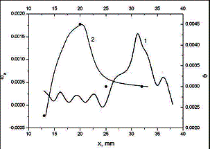 b)
b) 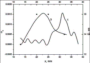
Fig. 1. The dependencies of block misorientation
(2) and size (3) on the distribution of local rotations (1)
Using the method of transmission electron
microscopy, structural investigation was performed for material sampled from
the area in which fracture was liable to occur. By and large, the characteristic
sizes and morphology of structural components are found to remain unchanged. However,
a fraction of subgrains would become non-equiaxial ones. It is also found that
most structural components are elongated along the extension axis. The extent
of non-equiaxiality is as high as 2.5. That subgrains and grains having sizes
0.1…0.4 μm account for 80% and nonequiaxial subgrains and
grains having sizes 0.6…0.9 μm, the remaining 20%. Of particular interest are the research results obtained
by the technique of atomic force microscopy in conjunction with the thin-foil method.
Thin-foils are generally employed for transmission electron microscopy
investigations. The surface relief of thin foil examined was found to contain
deep equiaxed cups (etch pits) having average size 0.14 μm as well as nonequiaxed ones having average size
0.19 μm (maximal diameter) and 0.09 μm (minimal diameter), i.e. the extent of
non-equiaxiality is ~2. It is noteworthy
that non-equiaxed cups had longitudinal axis aligned along the sample loading
axis. These results were matched with the data obtained by transmission
electron microscopy. There is good reason to think that the appearance of deep
cups on the foil surface is due to chemical polishing, which caused etching out
of structural components of ultrafine grain titanium. The granular structure of
material is recognizable from the partially etched-out grain boundaries. This
observational result can be used for certification of materials having nano-
and submicrocrystalline structure.
The
co-authors are thankful to the Russian Foundation of Fundamental Research for partial
financial support of the investigations which were performed in the frame of
Grant No. 14-08-00299.
References
[1] G.V. Shlyakhova,
A.Yu Eroshenko, V.I. Danilov, Yu.P. Sharkeev, A.I. Tolmachyov,
Microstructure and
fracture features of ultra-fine-grained titanium VT1-0 produced by abc-pressing method, Deformation and Fracture of
Materials 9 (2012) 24-28.
[2] L.B. Zuev, V.I.
Danilov, S.A. Barannikova, V.V. Gorbatenko, Autowave model of localized plastic
flow of solids, Phys. Wave Phenom. 17 (2009) 66-75.
[3]. G. F. Kuznetsov,
X-ray topographic identification and measurement of plastic strains and elastic
stresses in single crystallites of polycrystalline diamond layers, Technical
Physics 48 (2003) 1546-1553.