AGE-SPECIFIC
PECULIARITIES OF THE IA AFFERENT INNERVATION OF HUMAN M. SOLEUS
A. Chelnokov
Velikiye Luki State Academy of Physical Education and
Sports, Velikiye Luki, Russia
The aim was to
study age-specific peculiarities of the afferent Ia innervation of human m.
soleus.
 The experiment was conducted on 24 males, 9 to
18 years old (9-12 year-old boys, 14-15 and 17-18 year-old teenagers). The
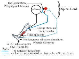
method of recording of Ia afferent presynaptic inhibition (PI) m. soleus under
homonymous vibrating stimulation at tendo calcaneus (N.P. Anissimova et al.,
1987, Figure 1) was used to evaluate the afferent input m. soleus.
The method is based on the evaluation of m. soleus H-reflex amplitude
suppression and its recovery in the aftereffect period. The more the m. soleus
H-reflex suppression, the higher the PI of the n. tibialis Ia afferent fibers.
The persons being tested laid down on a special couch, in the back-lying
position, with their legs outstretched and their feet drooping freely over the
couch edge. A DC vibrator motor DMP-30-H1-01 with an eccentric was used. The
vibrator was attached to the right shin above t. calcaneus with a special
rubber band. Moderate vibration was applied, for selective activation of the Ia
afferent fibers of m. soleus. The stimulation frequency was 65 Hz, the
oscillation amplitude was 0.25 mm irrespective of the vibrator pressing force.
The m. soleus H-reflex was recorded during a 60-second period before the
vibration stimulation, during a 30-second period of vibration stimulation, and
during a 60-second aftereffect period. The interval between the testing stimuli
applied to n. tibialis was 10 seconds. The m. soleus biopotentials were taken
using bipolar electrodes. Signals from the biopotential amplifier output were
supplied through an analog-to-digital converter to one of the PC ports. For
processing, the Myo software (ANO Vozvrascheniye, St. Petersburg, Russia, 2003)
was used.
The experiment was conducted on 24 males, 9 to
18 years old (9-12 year-old boys, 14-15 and 17-18 year-old teenagers). The
method of recording of Ia afferent presynaptic inhibition (PI) m. soleus under
homonymous vibrating stimulation at tendo calcaneus (N.P. Anissimova et al.,
1987, Figure 1) was used to evaluate the afferent input m. soleus.
The method is based on the evaluation of m. soleus H-reflex amplitude
suppression and its recovery in the aftereffect period. The more the m. soleus
H-reflex suppression, the higher the PI of the n. tibialis Ia afferent fibers.
The persons being tested laid down on a special couch, in the back-lying
position, with their legs outstretched and their feet drooping freely over the
couch edge. A DC vibrator motor DMP-30-H1-01 with an eccentric was used. The
vibrator was attached to the right shin above t. calcaneus with a special
rubber band. Moderate vibration was applied, for selective activation of the Ia
afferent fibers of m. soleus. The stimulation frequency was 65 Hz, the
oscillation amplitude was 0.25 mm irrespective of the vibrator pressing force.
The m. soleus H-reflex was recorded during a 60-second period before the
vibration stimulation, during a 30-second period of vibration stimulation, and
during a 60-second aftereffect period. The interval between the testing stimuli
applied to n. tibialis was 10 seconds. The m. soleus biopotentials were taken
using bipolar electrodes. Signals from the biopotential amplifier output were
supplied through an analog-to-digital converter to one of the PC ports. For
processing, the Myo software (ANO Vozvrascheniye, St. Petersburg, Russia, 2003)
was used.
Figure 1. Vibration
stimulation method of determining presynaptic inhibition of Ia fibers (N. P. Anissimova et al.,
1987).
Different human ontogenesis periods are described with different
intensities of the PI of m. soleus Ia afferent fibers. The research results
have shown that, under homonymous vibration stimulation of t. calcaneus, the PI
of m. soleus afferent fibers is considerably higher in 17-18 year-old teenagers
than in 9-12 year-old boys and 14-15 year-old teenagers (Figure 2).
Analysis of Figure 2 shows
that the same tendency in dynamics of m. soleus H-reflex amplitude
suppression under vibration stimulation
of t. calcaneus is observed in all age groups. The maximum PI values are
observed at the 30th second of vibration stimulation of t. calcaneus, while the minimum values, as
compared to rest, are observed at the 1st second of vibration.
Thus, at
the 30th second of vibration stimulation, the m. soleus H-reflex
decrease, as compared to rest, was 1.10 mV (89.02%) in 9-12 year-old children,
2.67 mV (78.96%) in 14-15 year-old teenagers and 0,75 mV (89,91%) in 17-18
year-old teenagers.
В А
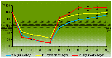
Figure 2. Dynamics of m.
soleus H-reflex amplitude during vibration stimulation of t. calcaneus (A) and aftereffect
period (B) in 9-12 year-old, 14-15 year-old teenagers boys and 17-18 year-old
teenagers.
The research results demonstrate different rates of recovery of PI of m.
soleus primary afferent fibers after vibration stimulation of t. calcaneus in
studying age groups. Thus, 17-18 year-old teenagers showed faster PI recovery
after the vibration stimulation as compared to 9-12 year-old boys and 14-15
year-old teenagers. An evidence of this is faster increase of m. soleus
H-reflex amplitude to the background values after vibration stimulation in
17-18 year-old teenagers than in other age groups. In 17-18 year-old teenagers,
the recovery of PI of the m. soleus Ia afferent fibers was observed at the 30th
second (113,92%) after the vibration end, while with 14-15 year-old boys, this
was observed at the 40th second (96,70%), while with 9-12 year-old
boys, this was observed at the 60th second (91,50%) (Figure 2).
In our opinion, the change of intensity of the PI of the m. soleus
primary afferent fibers, under homonymous vibration stimulation, is caused by
the development of reflectory functions of the neuromotor apparatus, which is
associated with the level of the morphofunctional maturation of its links and
their anatomic changes in the process of the human age-related development.