*99362*
K.B.Djusupov,
V.O.Kenbayev
Diagnostics and
treatment odontogenic mediastinitis
Shymkensky city hospital
of the first help
Introduction.
Sharp
odontogenic the infection is one of actual problems of modern surgical
stomatology.
Last
years the increase in number sick sharp odontogenic is marked by inflammatory
diseases, the heavy, progressing current, becoming complicated by sharp
respiratory insufficiency, mediastinitis, meningoentsifalitom and other
intracranial inflammatory processes, a sepsis, septic shock (1,2) is quite
often observed.
Despite
the certain successes reached in treatment sharp odontogenic of inflammatory
diseases and their complications, a lethality continues to remain high that
testifies to necessity of early diagnostics, forecasting of a current and
effective treatment.
Change
of a clinical picture of disease, especially in the beginning of its
development that creates diagnostic difficulties (3) is quite often noticed.
Abundantly
clear that increase and increase in weight of a current of inflammatory
diseases have led to considerable growth of time invalidity, and in some cases
to physical inability of an analyzed category of patients.
Thus,
the considered problem has not only medical, but also important social value.
According
to different authors, frequency of lifetime diagnostics mediastinitis makes
20,5% - 50%, and now disease diagnostics continues to remain one of difficult
solved problems (4).
Complexity
of early diagnostics odontogenic mediastinitis speaks absence of symptoms,
pathognomonic for mediastinitis early stages of its development, complicated
differential diagnostics of phlegmons of maxillofacial area, a neck and
odontogenic mediastinitis and its treatments.
The
purpose of this study was to examine pathognomonic signs in odontogenic
mediastinitis, the effect of systemic enzyme therapy on the course of the
inflammatory process.
Materials and Methods.
We
archive the data analyzed SHGBSMP over the past 10 years. It was found in 12
patients with odontogenic mediastinitis, at the age of 31 to 52 years.
All patients, depending on the type of
treatment were divided into 2 groups.
Group
1 consisted of 5 patients who were treated with traditional methods without the
use of systemic enzyme therapy.
Group 2 consisted of 7 patients treated with
systemic enzyme.
In particular, "Wobenzym" was used
for oral administration of 30 tablets per day. For local application of the
enzyme was used the following technique: after surgical drainage of purulent
focus and source of necrotic tissue, wound treated with antiseptics (hydrogen
peroxide, chlorhexidine, etc.), then watered it with a solution of this enzyme.
Enzyme solution was prepared at the rate of 25-50 mg "Vobenzima" to
5-8 ml of isotonic solution. In the purulent wound initially injected solution
from the syringe. This irrigation enables better contact with the enzyme
tissue.For large wounds, irrigation was carried out with solutions of the
enzymes were injected simultaneously gauze sponges soaked in solutions of the
same enzymes. Superimposed on the wound aseptic bandage with hypertonic saline.
Results.
For
odontogenic mediastinitis in late diagnosis is characterized by its progressive
course with lightning-like spread of purulent-necrotic process in the
background of impaired immunity to all parts of the mediastinum to the
development of polyorganic and hemodynamic disorders, mental disorders,which is
typical for the clinic of infectious-toxic shock.
In recent decades, the development of
techniques of cultivation under anaerobic conditions in the etiology of
odontogenic mediastinitis clarify the role of obligate anaerobic microorganisms
inhabiting the oral mucosa (5).That anaerobic bacteria are nesporogennye
etiologic agent of odontogenic mediastinitis. Synergy aerobes and anaerobes
leads to increased virulence of microorganisms and promotes an aggressive
course of the inflammatory process, the rapid melting of the tissue and severe
intoxication, aggravated by the lack of timely laboratory confirmation.
In
16% of cases the disease has developed against a background of relative
physical health, 84% of process took place in the presence of any underlying
disease.
Most
other diseases encountered chronic alcoholism (12% of cases), cardiovascular
failure (11%), diabetes (9%), renal-hepatic failure (8.1%), etc.
Risk
of developing acute inflammatory process in the mediastinum consisted of
patients with lung diseases (asthma, tuberculosis, chronic obstructive
bronchitis), gastrointestinal tract (chronic gastritis, gastric ulcer and duodenal
ulcer) blood (iron deficiency anemia,chronic lymphocytic leukemia).
Mediastinitis is characterized by a syndrome
comprising the triad, each of which is due to an independent pathogenetic
mechanism.
The
first symptom - pain involves a group of symptoms characterized by increasing
pain in the retrosternal space, which is enhanced by crowding the head (symptom
Gerke), palpation, stroking upwards or delaying neurovascular neck (a symptom
of Smith),swallowing and cough.
Coughing
symptom characteristic of odontogenic mediastinitis as a consequence of edema
floor of the mouth, soft palate and peripharyngeal space in the development of
phlegmon of the locations, which is always accompanied by irritation of the
tongue.
With
the development of acute inflammation in the mediastinum appear
symptom-Shcherbo Ravitch, characterized by retraction of the skin in the area
of the jugular depression
during inspiration, and paravertebral symptom Steinberg - the appearance of
rigidity of muscles.
On
the possibility of acute inflammation in the mediastinum shows
symptom-Rutenburga Revutskiy characterized by the appearance of pain in the
chest with displacement of the trachea.
In
the later stages of development of mediastinitis, if the total defeat of the
mediastinum, there may be a symptom of compression Popov - strengthening of
chest pain and the appearance of cough reflex with effleurage of the calcaneus
with extended lower limbs in the patient lying down.
In
addition, there may be a positive phrenic symptom - pain in the hypochondrium
and muscle tension anterior abdominal wall.
At
the rear of mediastinitis note of pain in the interscapular irradiation or
epigastralnuto field and gain the slightest strain, and with pressure on the
spinous processes of the vertebrae, especially the 5th baby.
All
patients had mediastinitis is defined sharp pain in the sternum and ribs. If
the subcutaneous tissue of the neck or chest is accumulation of gas, revealed
crepitations symptom. The development of pain determines the forced position of
the patient in bed, as an attempt to straighten causes increased pain in the
back, chest and in the throat.
The second group of symptoms is determined by
increasing intoxication.
The
patient is disturbed consciousness, somnolence, areactivity, apathy, delirium.
Sometimes, in severe cases, delirium develops intoxication, which is showing
signs of aggression.
Less commonly observed euphoria, quickly
giving way to loss of consciousness manifestation of the terminal state.
The
third group of symptoms is determined by the compression of blood vessels and
nerves. In many patients the superior vena cava syndrome, manifested by
swelling of the upper torso, neck and face, increased subcutaneous veins. This
is accompanied by increased headache, increasing tinnitus, cyanosis of the
facial skin.
Compression
of large vessels and nerves, leading to dysfunction of internal organs, and
compression and irritation of the purulent exudate of the vagus nerves causes
heart rhythm disturbances, bradycardia, bronchospasm. A number of patients we
observed sinus tachycardia, atrial fibrillation. As the relief of the inflammatory
process in the mediastinum state infarction improved.
On
the involvement of an acute inflammatory process of sympathetic trunk shows
symptom Horner.
Symptoms of irritation of the phrenic nerve
is a hiccup. Due to compression of the phrenic nerve arises diaphragmatic
paralysis, which can lead to respiratory failure.
Among the very important and severe symptoms
can include effects of compression of the trachea, main bronchi and esophagus.
In such cases, the clinical picture becomes
very severe mediastinitis.In addition to the compression of these organs, their
displacement occurs, and the destruction of their walls.
Compression
of large vessels and nerves, causing resorption of toxins and decomposition
products of tissue, which, according to clinicians, enhances cardiovascular
function disorders and respiratory systems.
Clinical
analysis of results of treatment of the second group showed that the most
pronounced therapeutic effect was obtained with local application
"Vobenzima" and administered orally in large doses.
Thus,
patients with the first group, where we used the traditional method of
treatment of the stabilization process of advancing to 7-8 per day. Share of
the mortality was 41.6%. Whereas in group 2, the stabilization process took
place for 4-5 days, the percentage of mortality was 14.2%.
To
illustrate typical observations give the following extract from the history of
the disease:
Patient M.D, 34 years old. Case history
number 8960, was admitted to hospital on the third day 20/10/09 from onset.
Complaints
of general weakness, headache, fever, sleep, appetite, dry mouth, difficulty
swallowing, breathing.
Locally defined abrupt swelling of the bottom
of the mouth, skin hyperemic and edematous.On palpation determined sharply
painful infiltrate without sharp boundaries in the submental and submandibular
regions on the left and right. Mouth opening is limited to 1.5 cm due to an
inflammatory contracture. Language is increased, overlaid with a purulent
coating.
On
admission the patient was determined in blood leukocytosis 28.9 x 10⁹
/ l, neutrophilia, toxic granulation of neutrophils, leukocyte shift to the
left, accelerated erythrocyte sedimentation rate 30 mm / h. The urine was
observed proteinuria, leukocyturia, cylindruria.
In
the analysis of biochemical parameters established hypoproteinemia 48 ± 1,8 g /
l, hyperglycemia, 6,84 ± 0,76 mmol / liter.
Chest
radiography in frontal projection possible to determine the extension of the
median shadow, blurring its outlines (Fig. 1).
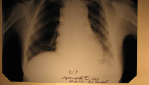
Figure
1. X-ray study. Patient ID number
Mukhamedova history 9980.
However,
we found that expanding the boundaries of the mediastinum and the
retropharyngeal space is far from the neck in all patients. It depends on the
mechanism of inflammation in the tissue of the mediastinum (Fig. 2).
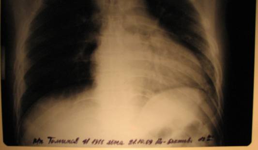
Figure
2. X-ray study.
Of
radiographic methods of investigation the most common X-ray of neck and
mediastinum in two projections, which must be done in the dynamics of every 2-3
days (Fig. 3).
For
the diagnosis of odontogenic mediastinitis performed X-ray examination of the
neck and lateral projections, which identifies the expansion of the shadow of
retropharyngeal space, the presence of gas in soft tissues and in
retrofaringealnom space.
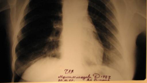
Figure
3. X-ray study of the dynamics. Patient
ID number Mukhamedova history 9980.
Clinical
diagnosis: "odontogenic phlegmons floor of the mouth. Odontogenic
mediastinitis. "
Under
general anesthesia, the incision is made on the angle of the mandible to the
angle. Then the wound bluntly extended covered in submaxillary bolast left and
right submental region, received up to 15 gnynogo discharge with bad odor.
Wound washed with antiseptic solutions (hydrogen peroxide, potassium
permanganate). After that, the wound irrigated with a solution from the syringe
by the enzyme "Wobenzym", and the wound was introduced rubber tube,
which is around zatamponirovana gauze, saturated solution of the enzyme
"Vobenzima." On the surface of the wound dressing was applied
antiseptic with hypertonic saline.
Cervical
access to the mediastinum, the proposed VI Razumovsky in 1899, is convenient
and malotravmatichen, allows for adequate disclosure of deep phlegmon of the
neck kletchatochnyh spaces, particularly retropharyngeal space.
Appointed general medication. A similar
purulent wound dressing, the patient was carried out on a daily basis.
After 4-5 days the patient's condition
improved and pain decreased.
In good condition the patient was discharged
from the hospital (02.11.09g) for outpatient treatment.
The
basis of treatment of odontogenic mediastinitis is prompt surgical
intervention, consisting in the disclosure under general anesthesia phlegmon
kletchatochnyh deep space neck and active drainage and sanitation foci of
chronic odontogenic infectionwhich caused inflammation. Severe condition of the
patient with mediastinitis can not be regarded as a contraindication to
surgery.
Active
surgical treatment of purulent diseases pathogenetically substantiated and
practically justified, because in reducing treatment time, achieving good
functional results and lower mortality rates (Figure 4).
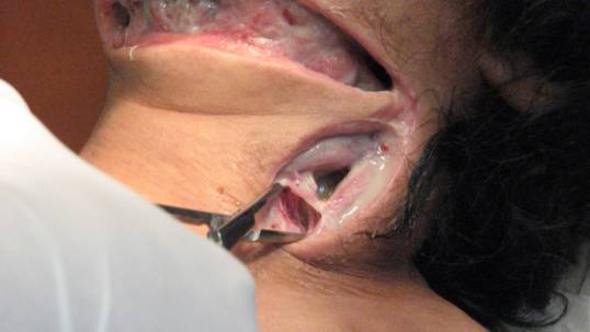
Figure
4. Patient ID number Mukhamedova history 9980.
Comprehensive
treatment program phlegmon deep neck spaces kletchatochnyh complicated by
mediastinitis contact, is to implement a pathogenetically based measures aimed
at suppressing the pathogen, the correction of hemodynamic and metabolic
disordersfight against intoxicationincrease of nonspecific resistance and
immunological reactivity. Remedial measures already after 3-4 days ensure
reduction of toxicity, body temperature, the patient feel better.
If not, there is reason to believe that the
outflow of pus is not enough, or you can think about the possibility of any
infectious and inflammatory complications.
The
outcome of odontogenic mediastinitis is in direct proportion to the length of
hospitalization. According to our observations in the delivery of patient care
within 3-4 days after the onset of primary tumor mortality from mediastinitis
was 31.3% at admission in a period of 4 to 6 nights - 41.2% in the period from
7 to 9 days - 51.7%.
On
admission patients at a later date adverse outcomes reported in 100% of cases.
In all cases, adverse events were detected in
sections of diffuse purulent or septic mediastinitis, purulent pericarditis,
pleurisy and pneumonia. With the development of mediastinitis seen against the
background of sepsis venous plethora of parenchymal organs: liver, spleen, and
kidneys.
Reference.
1.
Egorova O.A Clinical features odontogenic mediastinitis due to the mechanism of
its development. Clinical trial, Abstract. dis .... candidate of Sciences. St.
Petersburg 2002;
2. V.A Kozlov acute hospital dental care. A:
Medicine 1998; 288.
3. Kozlov VA, Egorov OA Odontogenic
mediastinitis. Clinical picture, diagnosis,treatment. St. Petersburg: MAPS
2002;
4. Oleinik I.I, Ponomarev A., Tsarev R.H,
Kurakin A. The species composition of associations of pathogens odontogenic
infection and prospects for cross antibiotics. Voen.med. Journal 1992;
5.Diaine B., Albertini M., Coussement A. Mediastinal extension of retro-pharyngeal
abscess. J Radiol 1992; 73: 4: 229-233.
6. Mevio E. Anaerobic cervical cellulilis: a
therapeutic approach. Acta Otorlii-nolaryngol Ital 1993; 13: 6: 525-536.