а) plaster
cast b)
schematic mo
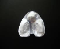
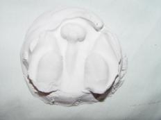
Fig. 1 – The general view of plaster cast (а) and its` schematic
image (b)
in patient who was delivered
to the hospital (1level)
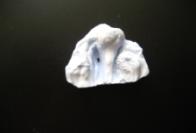
The second
level of an intermaxillary bone and the
side fragments of the upper jaw were found in 26 patients. They had a
transverse dimension of the intermaxillary bone -20 mm, protrusion - from 8 mm
up to 15 mm, the angle of deviation from the median line - up to 10 degrees, displacement from the vertical plane - from 2 up
to 4 mm. Distance between the front edges of the side fragments of the alveolar
process of the upper jaw was up to 15 mm. The width of the defect on the border
of hard and soft palate was 19,3 ± 1,0 mm (Fig. 2a, b).

а) plaster cast
b) schematic model image
Fig. 2 – The general view of
plaster cast (а) and its` schematic image (b)
in patient who was delivered to the hospital (2 level).
The third level of an
intermaxillary bone deformation and the side fragments of the upper jaw
were found in 23 patients. They had a transverse dimension of the
intermaxillary bone up to 25 mm, protrusion -from 16 mm and more, the angle of
deviation from the middle line - from 11 degrees and more, the displacement by
the vertical plane –from 5 mm and more. The distance between the front edges of
the side fragments of the alveolar process of the upper jaw was up to 25 mm.
The width of the defect on the border of hard and soft palate was 22,7 ± 1,0 mm
(fig. 3a, b).
а) plaster cast b) schematic model image
Fig.3 – The general view of plaster cast (а) and its` schematic
image (b)
in patient who was
delivered to the hospital (3 level).
The division of patients
with congenital bilateral cleft lip and palate into 3 groups before the treatment
had a practical importance, which was taken into account when planning the
treatment. Usually, we took to the treatment patients with the first and partially
with second part of deformation degree of Intermaxillary Bone, by using of removable orthodontic appliances
[7]. In the case of the intermaxillary
bones deformations of the third degree the presurgical preparation of patients
was performed by using of innovative technologies (non-removable devices, fixed
microimplants, or use of microimplants).
Clinical
application of microimplants. All patients (23 children) with congenital bilateral
cleft lip and palate with third degree of
intermaxillary bone deformation
the presurgical orthodontic treatment was executed by using of
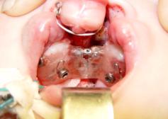
microimplants. At the begining, the microimplants have been used in 6 patients
for intraosseous fixation of
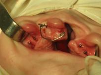
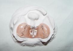
supramaxillary non-removable orthodontic appliances (Fig. 4). Others 17 children
with such pathology used the microimplants as supporting elements of
orthodontic appliances (Fig.5).
Presurgical orthodontic preparation of patients was
carried out in several stages. At the beginning the plaster casts of the upper jaw have been taken from children at
the children dental clinic. After that,
the models of jaws were under
preparation. Analyzing the results data
of diagnostic models of the upper jaw and the parameters of the
computer-tomographic study the place of installation microimplants was
determined.
Within
children's clinic conditions of maxillo-facial surgery under the general
anesthesia, 6 patients have been installed by supramaxillary non-removable
devices that are fixed to the bone by using of microimplants (Figure). For this
purpose the holes in the projection angle of 45 °C to the top of the comb of
the alveolar process were prepared beforehand on the fixed fragments (two side
and maxillary bones). At the same time the both sides fragments were connected
by a V-shaped orthodontic screw for expansion of the side fragments of the
upper jaw. This structure of the apparatus used for the expansion of the side
fragments of the upper jaw and for repositioning the intermaxillary bone in the
correct position, forming the alveolar arch.
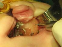
4 5
Fig. 4 -patient А-v С. , 4 months., bilateral cleft lip and palate after fixation supramaxillary non-removable device to the upper
jaw fragments and to the intermandibular bone by
microimplants.
Fig. 5 -
patient V-yev, 5 months., bilateral
congenital cleft lip and palate after installation of the microimplants to the
upper jaw fragments and to the intermandibular bone
for orthodontic treatment.
The next step was to adapt the child to the
apparatus and the adjustment
of its
feeding, training the mother for hygiene care of oral cavity in child and
apparatus.The location of microimplants in the side fragments of the upper jaw
was monitored by panoramic radiographs. Depending on the children health
status the children were discharged
from the hospital on 6-7 days after surgery under the supervision of
orthodontist.
After the child's adaptation to the device
the screw is activated by 0.5 mm once in two days, with simultaneous activation
of an elastic traction on one link in the three days. Taking into account the
continued growth of divided alveolar process of upper jaw at length after
cheiloplasty the fragments cleft of the upper jaw shouldn`t be completely locked, leave the diastasis
between them on the width of the temporary tooth. The duration of an active
period ranged from 20 up to 30 days. One of the important step is the retentional
period, which ranged from 15 up to 30 days. After the end of retentional period
the device was removed, and primary cheiloplasty on both sides has been
conducted immediately.
Last time we use microimplants
in order to transfer separate fragments of the upper jaw and shape them into dental arch in account with severity degree of an
intermaxillary bone and side fragments of the alveolar processes of the upper
jaw. Such early orthodontic treatment is one of the few samples of an innovative
technology using during presurgical treatment of the children with
congenital bilateral cleft lip and palate before surgical intervention.
If the microimplants were used without using of supramaxillary orthodontic
apparatus the moving of the jaw fragments and the normalization of dentoalveolar arch have been occured during 1 month after the
start of orthodontic treatment.
Thus, the
deformations study of the alveolar and palatal processes of the intermaxillary bone and
palate in children with congenital bilateral cleft lip and palate has allowed
us to approach differentially to the various methods of presurgical preparation
of children with such pathology. The literature data and our own research are allowed
to emphasize if more time passes before the surgery without the orthopedic and
orthodontic treatment, then the deformation of the intermaxillary bone and vomer will be exacerbated. It is occurs because of the influence of tongue and nipple, which is
especially noticeable with very severe
deformation of the intermaxillary bone and vomer. So which means that in
children with congenital bilateral cleft lip and palate the deformity of the intermaxillary
bone is increased by age, and it`s depends on the initial indexes of the disease
severity (mild, mean, severe), and also depends on the ways of children
preparation for the surgery in the presurgical period.
The application of the microimplants within presurgical preparation of the patients with cleft lip (cheiloschisis) and cleft of palate plays the main role in
the surgical rehabilitation and it helps to the surgeons to make primary
surgery without any difficulties and also it has a positive effect on the
healing of lips tissues.
For comparison characteristic
of the results of the different methods of presurgilal preparation of the children with congenital cleft lip
and palate the anthropometric studies of the
jaws were done. (Table).
Table
- Anthropometric data received from the jaws models of the children with bilateral congenital cleft lip and palate,
prepared for the surgery by different methods.
|
Age periods |
Distribution
of the children by treatment methods |
||||||||
|
Without
presurgical orthodontic preparation (p=45) |
Treated
patients by T.V. Sharova`s method (p=48) |
The
patients treated by using of microimplants
(p=28) |
|||||||
|
Side of
displacement of the intermaxillary bone |
On the side from the intermaxillary bone displacement |
Total |
On the
side of the intermaxillary bone displacement |
On the side
from the intermaxillary
bone displacement |
Total |
On the
side of the intermaxillary bone displacement |
On the
side from the intermaxillary
bone displacement |
Total |
|
|
1 month |
8,5 ±0,52 |
10,9 ±0,42 |
19,4 ±0,47 |
10,5 ±0,83 |
11,5 ±0,65 |
22,0 ±0,74 |
8,4 ±0,92 |
11,6 ±0,87 |
20,0 ±0,71 |
|
3 months |
9,4 ±0,95 |
12,1 ±0,62 |
21,5 ±0,81* |
8,8 ±0,70 |
10,9 ±0,88 |
19,7 ±0,79* |
8,3 ±0,93 |
11,5 ±0,86 |
20,3 ±0,89 |
|
6 months |
9,8 ±0,92 |
12,7 ±0,74 |
22,5 ±0,83* |
6,4 ±0,57 |
7,6 ±0,62 |
14,0 ±0,59* |
1,2 ±0,27 |
1,4 ±0,32 |
2,6 ±0,29* |
|
12 months |
8,1 ±0,84 |
10,7 ±0,88 |
18,8 ±0,86 |
3,5 ±0,39 |
5,5 ±0,24 |
9,0 ±0,31* |
0,9 ±0,17 |
1,1 ±0,18 |
1,0 ±0,18* |
* The differences statistically are significant in
comparison with indices, received from newborn of 1 month (Р<0,05).
As shown in the table, in patients of the comparable group with the growth
of a child with congenital bilateral cleft lip and palate the defects between
the intermaxillary bones and side fragments of the upper jaw increase and reach
its maximum at 6 months. After cheiloplasty it was observed statistically unauthentic
decrease of the bone defect.
In patients who were prepared for surgery with removable orthodontic devices, the bone
defects statistically and significantly decrease from 22,0 ± 0,74 mm up to 9,0 ± 0,31 mm, but the full butt contact
between the intermaxillary bone
and side fragments of the upper jaw was not occurred.
After
executed presurgical preparation with using of non-removable supramaxillary devices fixed
by microimplants, and also by microimplants it was reached the full frontal
contact between the intermaxillary bone and fragments of the upper jaw, which
was confirmed by static treatment of the material (P ˂ 0,05).
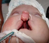
For illustration we present the stages of complex
treatment of the S.Yu-va patient (Fig. 6), who came to the clinic at 3 months
of age. After third degree determination of the intermaxillary bone the presurgical
patient preparation was carried out by using of the innovative technologies
(non-removable supramaxillary orthodontic device, fixed by microimplants). Bilateral
cheiloplasty was made at the age of 6 months and sparing palatoplasty-at the
age of 1.5 years.
А Б
В Г
Д Е
Ж З
И
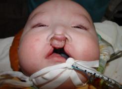
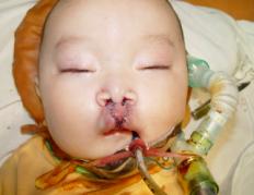
Fig. 6 - Patient S.Yu-va, 3 months.,
diagnosis: Congenital bilateral cleft lip and palate (3 degree of the intermaxillary bone deformation):
А) Patient`s appearance when
delivered to the hospital,
B)
Plaster cast of the frgaments of the upper jaw before treatment,
C)
The fitting of supramaxillary orthopaedic device on the plaster cast,
D) The fixation of supramaxillary orthopaedic device by microimplants to the jaw
fragments,
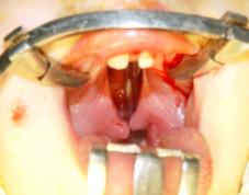
E) Intermaxillary bone status in patient at the age of 6 months,
F)
The patient state after bilateral cheiloplasty,
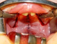
G) Palate tissues status in patient
at the age of 1,5 month before surgery,
I) Palate tissues status in patient
at the age of 1,5 month after palatoplasty,
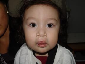
J) Patient`s appearance S. Yu-va at
the age of 2 years.
Summary. The modern design of
devices allows to reduce presurgical
preparation, to enter into the design active elements, to normalize the location
of intermaxillary bone and the upper
jaw form, and without special difficulties to produce a primary surgery of cheiloplasty,
after that the palatoplasty, accelerate the timing of complex rehabilitation of
patients with such complicated pathology of maxillofacial area as bilateral congenital cleft lip and palate with good aesthetic results.
Literature.
1. A.S. Artyushkevich, V. I. Phillipenko, L.S. Krishtopenko
and others: New in the treatment of the cleft lip and . // Materials of IV congress
of the dentists of Byelorussia: «Arrangement, prophylaxis, new technologies
and rehabilitation in dentistry». – Vitebsk, 2000. – P. 342–344.
2. E.S. Katasonova Substantiation of using new technologies at
early growth of holiatry of the children with congenital bilateral
cleft lip and palate: author's abstract
of candidate dissertation – Almaty,
2010. – P. 20.
3.
Turley P.K., Kean C., Schur J. et all.
Orthodontic force application to titanium endosseous implants. // Angle
Orthod. – 1988. - Vol.58, N 2. – P.
151-162.
4. Gray JB, Smith R. Transitional implants for orthodontic
anchorage. // J Clin Orthod.- 2000.- Vol. 34, N 11.- P. 659-666.
5. S.I. Blokhina, G.V. Dolgopolova, Medical and social
rehabilitation of the children with congenital
cleft lip and //Dentistry and baby`s health: thesis report of 1-st
Republic conference.– М., 1996. – P. 20.
6.
Millard D.R. Improved primary surgical and dental treatment of clefts / D.R. Millard,
R.A. Latham // Plast. Reconstr. Surg. - 1990 - Vol. 86. - P. 856 - 871.
7. T.V. Sharova, G. I. Rogozhnikov Orthopeadic dentistry of infancy. – М.: Medicine, 1991. – 288 P.