Мedicine/7.
I.V. Rodionov, dr. tech. sciences, professor
FSBEI HPE «Saratov state technical
university of a name Gagarin Yu.A.», Saratov, Russia
Article is prepared with support the grant President
of the Russian Federation
Influence of superficial structure implants on
character of their interaction with the bone fabric
Аbstract
In
article influence microstructure of a surface materials implantation appointments
to character of contact interaction with a bone fabric is considered. The
role various kinds of surfaces medical
implants on efficiency of osteointegration processes and durability fastening
of products in a bone fabric is defined. It is shown that the best conditions
for osteointegration implants provide porous biocompatible coverings with heterogeneous
morphology.
Keywords: medical implants, biocompatible materials and coverings, a surface
microstructure, osteointegration.
Аннотация
В
статье рассмотрено влияние микроструктуры поверхности материалов имплантационного
назначения на характер контактного взаимодействия с костной тканью. Определена
роль различных видов поверхностей медицинских имплантатов на эффективность остеоинтеграционных
процессов и прочность закрепления изделий в костной ткани. Показано, что
наилучшие условия для остеоинтеграции имплантатов обеспечивают пористые
биосовместимые покрытия с гетерогенной морфологией.
Ключевые слова: медицинские
имплантаты, биосовместимые материалы и покрытия, микроструктура поверхности,
остеоинтеграция.
An
important and topical issue of effective use of medical metal implants in
maxillofacial surgery, traumatology and orthopedics is the informed choice of
optimal parameters of the surface structure of implanted materials to ensure
their lasting relationship with the surrounding bone tissue [1, 2]. This
relationship can be achieved mainly due to macro- and mikrointegration
interaction of implantable medical-technical structures to the bone. It is
necessary that the functional intraosseous implant surface had a high level of
biocompatibility and pronounced heterogeneous structure of the large number of
open pores, whose size should provide a normal bone cell penetration, followed
by overgrowth of the entire surface of the bone regenerate.
Оptimization
of osteointegration processes in
terms of metal implants is achieved by stimulation of reparative osteogenesis
and revitalization of the bone cell structures in the surrounding area of the
implant. These conditions are provided in the first place, osteoconductive
properties of materials determined by their phase-structural state and the
nature of the surface morphology [3].
The role of the surface microrelief in the
manifestation of material osteoconductive implant is confirmed by numerous
experimental data indicating the influence of surface microgeometry implantable
products for the mechanisms of interaction with bone tissue and the nature of
the relationship with it [3-5].
In this paper we consider the influence of type of
surface structures of metal implants in the process of osseointegration and
healing in the body.
So, it seems quite obvious that the use of a smooth
surface can not create a strong contact interaction of implants with bone due
to lack of the possibility of a small osteointegration
process and the real contact area of this surface with the surrounding tissue.
For example, the surface topography obtained by turning the metal implant has a
poorly defined unidirectional with no signs of morphological heterogeneity
(fig. 1). Surface with such a structure can not ensure effective communication
with the implant and bone tissue strong enough to consolidate the bone. Numerous
clinical studies on laboratory animals show the uselessness of such implants in
the treatment of various bone pathologies as maxillofacial department and the
musculoskeletal system, due to lack of flow around smooth implants processes of
active bone formation. Observed only in the presence of various degrees of
fibrous tissue, greatly complicating engraftment implants.
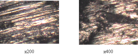
Fig. 1. The
surface of the implant from stainless steel (12X18H9T) with a smooth relief after turning processing
Giving a smooth metal surface of a high degree
of roughness greatly enhances its osteoconductive ability and integration
interaction with bone structures. Surface roughness determine the nature of its
physical and mechanical interaction with the surrounding tissue and create the
necessary conditions for the germination of bone cells in mikrohollows
different shapes and depths to form a relatively strong relationship with the
implant tissue. Therefore, the formation of a high degree of surface roughness
of implants received much attention. In addition, the initial roughness of the
implants can significantly improve the adhesion and porosity of the surface specially
applied bioactive coatings due to their strong mechanical bond to the substrate
particles and reproduction (copying) of the microrelief surface of the base
metal coating thin layers.
Materials developed rough surface is
characterized by a high level of energy has a definite influence on the degree
of adsorption of proteins from contacting biological media. This factor is an
important component of the initial implant healing process, because precedes
the subsequent cell proliferation and differentiation [3]. Due to the increased
specific surface area of rough implant materials increases and the
concentration on these adhesive proteins of the extracellular matrix and cytoplasm,
which leads to an accelerated accession to the surface of cells. At the same
time with a rough surface interacts significantly greater number of cells in
comparison with a smooth surface, so the proliferation, differentiation and
extracellular matrix synthesis occur faster on the rough structures of
materials.
The relief of a rough surface is characterized
by the presence of macro- and microscopic irregularities in the presence of
large protruding particles and deep depressions, and also of difficult focused
profile elements (fig. 2). Such a structure due to the high heterogeneity and
is capable of effective interaction of implants with bone tissue. This
heterogeneous structure is favorable for the occurrence of osseointegration and
may contribute to a strong biotechnical system «implant – the surrounding
bone».
However, creating the best conditions for the
occurrence of osteointegration processes
provide the оpenly porous biocompatible surface with the magnitude of the total open
porosity at the level of 30-60% and pore size of 20-200 microns. Such surfaces
are stimulated reparative osteogenesis and have high osteoconductive properties.
The large number of pores leads to a substantial increase in the specific surface,
contributes to the increased number of adsorption of adhesive proteins, accelerates
the cellular mechanisms of migration and transport biochemical systems,
creating optimum conditions for the formation of new bone tissue.
Openly porous systems at the expense of
maintenance high sorption to activity promote an intensification
of bone growth factors, that is
osteostimulation, causing accelerated bone formation. Such highly openly
porous surfaces are formed mainly by applying a metal implants bioactive coatings
based on biodegradable materials – this is usually calcium-phosphate ceramics
types (hydroxyapatite, ftorhydroxyapatite and oth.), have expressed
osteoconductive characteristics (fig. 3).
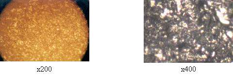
Fig. 2. The
surface of the implant of a titanium alloy ВT6 with rough terrain
after sandblasting abrasive corundum dispersion of 450 microns
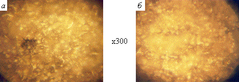
Fig.
3. Surface structure of bioactive calcium phosphate powder coatings on implants
made of titanium ВT1-0: a – hydroxyapatite covering, b
– ftorhydroxyapatite covering
However, the technical and economic point of
view calcium-phosphatic coverings is more expedient for using on implants
constant or long functioning, such as stomatologic inside maxillary lamellar,
conic and cylindrical bearing support of fixed tooth artificial limbs, and also
orthopedic osteoclamps of devices an external osteosynthesis, introduced in
various bone segments in a period of 7-9 months or more. In cases of temporary
implants with a term of operation of several weeks to 2-3 months is more
efficient use of porous coatings on the basis of carbon, bioglasses, oxides biologically
inert metals, polymeric composites (fig. 4). Coatings of these biocompatible
materials, first of all, can provide a solid relationship with the surrounding
bone tissue ingrowth through the pores of the surface microrelief and deepening
in developing a secure fixing of implants in bone, secondly, to ensure
subsequent atraumatic removal of implants from the bone structures of the body
parts at the expense of penetration of the bone only to a certain depth then
controlled by the technological regime of the coating.
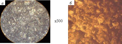
Fig. 4. The surface structure of oxide biocompatible coatings on metal implants:
a – air thermal oxide coating on the stainless steel 12X18H9T, b
– steam thermal oxide coating on titanium alloy ВT16 [6, 7]
Between the coated implant and biological media
is biophysical and biochemical interaction with the formation and adhesion to
the surface of the protein structures of the growth and penetration into the
grooves, as well as the pores of the surface relief in a certain degradation of
the material. As a result of this interaction is a durable implant
bioengineering system «implant – tissue surrounding the» high efficiency
operation. At the same time on the formation of osteoconductive properties and
bioactivity is greatly affected by the morphological heterogeneity of the
implant surface, defined set of indicators of roughness parameters as well as
projections and recesses, including the pores. Therefore, investigation of
surface microgeometry implant coatings is an important milestone in the
development and establishment of modern medical implants for reconstructive
surgery of different directions.
Thus, the increased effectiveness of the exhibit metal implants with
porous functional and morphologic development of coating materials is not only
compatible with biological structures, but also providing stimulate growth of
bone tissue, followed by rapid osseointegration.
References
1. Корж Н.А. Имплантационные материалы и остеогенез. Роль
биологической фиксации и остеоинтеграции в реконструкции кости / Н.А. Корж,
Л.А. Кладченко, С.В. Малышкина и др. // Ортопедия, травматология и протезирование.
2005. №4. с. 118-127.
2. Корж Н.А. Имплантационные материалы и остеогенез. Роль
индукции и кондукции в остеогенезе / Н.А. Корж, В.А. Радченко, Л.А. Кладченко,
С.В. Малышкина // Ортопедия, травматология и протезирование. 2003. №2. с.
150-157.
3. Корж Н.А. Имплантационные материалы и остеогенез. Роль
оптимизации и стимуляции в реконструкции кости / Н.А. Корж, Л.А. Кладченко,
С.В. Малышкина // Ортопедия, травматология и протезирование. 2008. №4. с. 5-14.
4.
Хлусов И.А. Генез костной ткани на поверхности имплантатов для остеосинтеза /
И.А. Хлусов, А.В. Карлов, И.В. Суходоло // Гений ортопедии. 2003. №3. с. 16-26.
5. Биосовместимые материалы: Учебное пособие / Под ред.
В.И. Севастьянова, М.П. Кирпичникова. М.: ООО «Медицинское информационное
агентство», 2011, 544 с.: ил.
6.
Патент РФ на изобретение №2412723. Способ получения оксидного биосовместимого
покрытия на чрескостных имплантатах из нержавеющей стали / Родионов И.В., Бутовский
К.Г., Анников В.В., Карпова А.И. Опубл. 27.02.2011.
7.
Патент РФ на изобретение № 2332239. Способ получения биосовместимого покрытия
на остеофиксаторах из титана / Родионов И.В., Бутовский К.Г., Бейдик О.В.,
Ткачева А.В. Опубл. 27.08.2008.