Prasol V.A., Troyan V.I., Ponomaryov A.O., Kupriyanova L.S.
Kharkiv National Medical University
(Ministry of Health of Ukraine) Pathomorphology Department
Immunological peculiarities of the wall of large subcutaneous vein in patients with varicose disease
Objective: To reveal immunohistochemical peculiarities of
the wall of large subcutaneous vein in patients with varicose disease.
Methods of research: Histological, immunohistochemical,
morphometry, statistical analysis.
Material: The controls were 4 cases of
autopsy of young male patients who died of different injuries. The study group
comprised 11 patients who were operated for varicose disease. In all cases a
segment of large superficial vein was investigated.
Results:
Microscopy of the vein walls of the study group demonstrated protrusions with
uneven thickening and consolidation of the wall. The vessel integrity was
preserved. Histological survey revealed that the wall of the vein in all cases
was presented by three layers: internal, medial, external. But the wall
thickness was changed. Thus, in the controls this parameter was 322.67±43.12х10‾³m, in the study group it was 490.27±18.65х10‾³m. In contrast to the controls, the changes of
myeloelastosis type were noted in the structure of the inner layer of the vein
wall in the study group. Massive growth of the connective tissue as well as
thickening and loosening of muscular fibers were observed in the structure of
the medial and external layers of the walls in of the study group (p. 1). Immunohistological method with Coons’
technique revealed the following peculiarities of collagen formation in the
walls of the veins of the investigated groups. In all observations type IV, I
and III collagens were present in the connective tissue. But the changes of fluorescence
intensity of these collagen types were noted.
The indices of collages
fluorescence intensity for main collagen types are presented in table 1.
Table 1
Type I, III, IV collagen fluorescence intensity in the connective tissue
of the vessel walls of the investigated groups (conventional units)
|
Group |
Type I collagen |
Type III collagen |
Type IV collagen |
|
Controls |
2.15±0.27 |
3.04±0.8 |
2.96±0.16 |
|
Varicose disease |
1.89±0.19 |
3.86±0.28* |
2.06±0.28* |
*р<0.05 (compared to the controls)
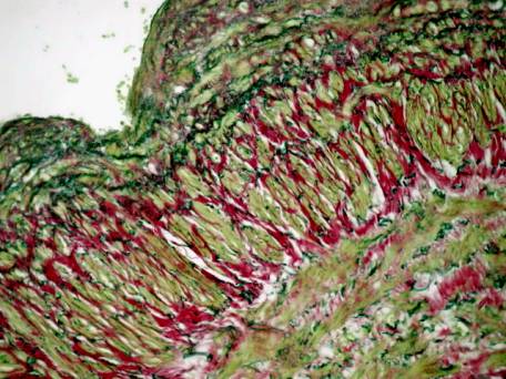
P. 1. Massive growth of the
connective tissue in the structure of the medial and external layers of the
walls in of the study group. Stained with Picrofucsin to van Geeson, x200.
The
data of the table suggest significant reduction of type IV and I collagens and
significant increase of type III collagen amount in the connective tissue of
the wall of large subcutaneous vein in patients with varicose disease (p. 2).
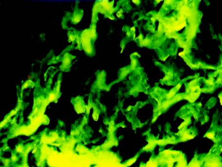
P. 2. Type III collagen in the
structure of the medial layer of the walls in of the study group.
Immunohistological method with Coons’ technique, x200.
Conclusion. The
complex investigation demonstrated prevailing sclerotic changes in main
components of the vein wall in patients with varicose disease which manifested
by changes in the structure of the connective tissue. The revealed
peculiarities of collagen formation can be a sign of connective tissue
dysplasia in patients with varicose disease.