CHANGE IN ACTIVITY OF CENTRAL AND AUTONOMOUS CIRCUITS OF
HEART RHYTHM REGULATION IN CASE OF DYSFUNCTION OF AIRWAYS
Doct of med. sciences,
prof. A.L. Isakadze, doct. of biol. sciences, prof. G.G. Eliava,
prof. T.G. Tsintsadze,
assoc. prof. L.S. Topuria
Tbilisi State Medical University
Georgian Technical
University
Annotation: Statistical analysis of heart rhythm
variability is widely used both in experimental and in clinical medicine.
In the given work, due to changes in respiratory function
there were studied the processes of rearrangement of performance level of blood
circulatory system, which are focused on maintenance of homeokinesis and which characterize
system mechanisms.
Research showed that during disorder of natural nasal
breathing under conditions of relative physiological rest takes place
displacement of balance (equilibrium) of central and autonomous circuits of
heart rhythm regulation, enhancement of slow periodicity and severity
(expressiveness) of stochastic aperiodic influences, decrease of index of
tension of regulatory systems, increase of index of functional status of heart.
Keywords: heart rhythm
variability, nasal breathing, central and autonomous circuits, heart rhythm regulation
I. Introduction
Mathematical description of physiological processes makes
possible not only quantitative, but also qualitative analysis of vital
processes in body and its systems under influence of different environmental
factors on it [1,7].
Mathematical analysis of heart rhythm is widely used both in
experimental and in clinical researches [1, 2, 7, 8].
Processes of rearrangement of performance level of blood
circulation system reflect not only the action of stress factors, but also characterize
internal and intersystem mechanisms focused on maintenance of
homeokinesis.
It is known that stress, which runs in three stages (alert
stage, resistance stage, exhaustion stage) is a unity of non-specific response,
which are based on neuroendocrinal and metabolic changes, which manifest
themselves as general adaptation syndrome (Fig. 1).
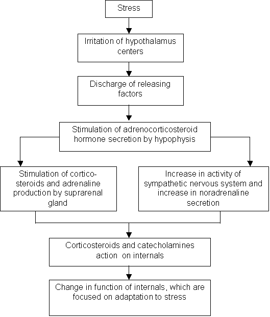
Fig. 1. Stress factor’s
action mechanisms
Homeokinesis abnormalities during stresses are preceded by
disorders in regulation of function, which emerge as a result of tension,
supertension and further depletion of adaptation mechanisms (Fig. 2).

Fig. 2. Influence of homeokinesis
function disorder on different indices
of vital functions

Fig. 3. Method of study of different aspects
of activity of regulatory systems
Regulation of body functions may be represented by three
interrelated systems – cerebral cortex, system of hormonal-humoral regulation
and vegetative nervous system (Fig. 3).
Different samples of RR intervals are used for acquisition of
information on various changes in body of healthy and sick persons depending on
assigned task.
Good correlation of mathematical-statistical indices of
heart rhythm and hemodynamics indices is obtained in patients having arterial
hypertension [2, 5, 6]. Obtained indices supplement clinical-postgenetic
features of formation and course of arterial hypertension. Received indices
testify the predominance of sympathetic activation of vegetative nervous system
among patients having arterial hypertension and the superiority of central
mechanism of regulation of heart rhythm variability over autonomous
regulations. Excessive activation of sympathetic-adrenal system and decreased
activity of parasympathetic regulation testify deadaptation processes in
hypertensive patients with disorder of regulation both at the level of
segmental (subcortical) and suprasegmental (nuclear) brain structures and
decreased influence of cortical inhibition on them.
Based on abovementioned we set a task of study of processes
of rearrangement of performance level of blood circulation system due to
changes in respiratory function.
II. Problem setting.
Heart rhythm variability of humans was studied in our work.
Study was conducted in healthy persons aged 19-20 years.
Goal of the work was a study of mathematical-statistical
indices of heart rhythm in case of disorder of natural nasal breathing. Persons
under test performed breathing under conditions of both nasal and mouth
breathing. In case of mouth breathing nasal breathing has been turned off using
soft nose clip. Study has been conducted under conditions of relative
physiological dormancy. Both nasal and mouth breathing have been performed in
seated position. For removal of stochastic fluctuations of measurement the persons
under test took a course of preliminary adaptation to mouth breathing. Method
of correlation rhythmography has been used for research. Registration of correlation
rhythmography has been conducted with the use of rhythmocardioscope
RKS-01.
Sample volume was 100 cardiocycles. Mode (Мо), mode amplitude (АМо) and variation range (ΔХ) have been determined. By
means of special program there have been established integral indices of heart
rhythm: index of tension (IT) of regulatory systems; index of functional status
(IFS).
III. Results.
Research results testify that at nasal breathing takes place
the change of heart rhythm variability. At mouth breathing occurs enhancement
of aperiodic stochastic influence on heart rhythm.
At nasal-mouth breathing, in comparison with nasal breathing
takes place elongation of longitudinal axis of correlation rhythmogram, i.e.
slow periodicity becomes more expressed.
Similar dynamics of correlation rhythmogram is also observed
in case of only mouth breathing: takes place elongation of not only longitudinal
axis of correlation rhythmogram but also transverse axis of correlation
rhythmogram.
Abovementioned changes in correlation rhythmogram obviously
demonstrate that mouth breathing promotes both the increase of aperiodic
stochastic influence on heart rhythm and enhancement of slow periodicity in
heart rhythm.
Analysis of such statistical parameters of heart rhythm as
the mode (Мо), mode amplitude (АМо) and variational range ΔХ, evidences that at mouth
breathing takes place enhancement of heart rhythm variability (Table 1).
Table 1. Change
in indices of variational pulsogram at nasal and mouth breathing
|
Indices |
Nasal breathing |
Mouth breathing |
Statistical indicator of confidence level |
|
Mode, Мо |
0,79 ± 0,08 |
0,73 ± 0,02 |
P < 0,05 |
|
Mode amplitude АМо |
37,5 ± 4,8 |
22,0 ± 4,1 |
P < 0,001 |
|
Variational
range |
0,135 ± 0,001 |
0,20 ± 0,0045 |
P < 0,05 |
Indices of variational pulsogram are statistically significantly
changed (р<0,5)
as follows (Table 1):
- value of mode amplitude is decreased;
- mode value is decreased;
- variational range is increased.
Index of tension of regulatory systems, which has negative
correlation with the index of functional status of heart, under conditions of
mouth breathing significantly decreases (p<0,05) by 26% in average (Fig. 4).
Index of functional status of heart increases by 45,8% in average (Fig. 5).
IT

Fig. 4. Change
in index of tension (IT) of regulatory systems at nasal (1)
and mouth
(2) breathing
IFS
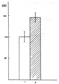
Fig. 5. Change in index of functional status
of heart at nasal (1)
and mouth (2) breathing
Abovementioned changes are in accordance with hemodynamic
researches.
According to literature data [1,2,5,6], slow waves are related to adaptive activity of
cardiovascular center.
Stochastic changes of venous inflow caused by the change in
functional status of cardio-vascular system at mouth breathing lead to
abovementioned changes of correlation rhythmogram. Changes in vegetative
homeokinesis are related to increased influence of both sympathetic (relative
gain of mode) and, predominantly, parasympathetic (enhancement of variational
range, decrease in index of tension of regulatory systems) divisions of
vegetative nervous system on sinoatrial node.
Change in vegetative homeokinesis may be explained by the
fact that takes place the disorder of balance
of central and autonomous circuits of heart rhythm regulation and, thereby,
character of heart rhythm regulation. It seems that these phenomena may be
explained by change of sensory input (afferent signalization) from airways
under conditions of different pattern of breathing [3, 4].
Research results give us an opportunity to suppose that extracardiac
effects, implemented from airways play significant role in multistage system of
heart rhythm control.
IV. Conclusions:
Under conditions of mouth breathing the influence of
autonomous circuit of heart rhythm regulation prevails.
1. Vegetative homeokinesis of heart rhythm regulation is
changed in case of disorder of natural nasal breathing. Balance of central and
autonomous circuits of heart rhythm regulation is disrupted.
2. Under conditions of mouth breathing is increased the
severity (expressiveness) of slow periodicity with simultaneous enhancement of stochastic
periodical effects.
3. Obtained data must be taken into account during
assessment of heart rhythm of patients with dysfunctions of upper respiratory
airways for delivery of adequate therapy.
References
1. Baevsky R.М. Mathematical methods of
heart rhythm analysis – М., 1961 (in Russian).
2. Baevsky R.М., Kirillov О.I., Kletskin S.M. Mathematical
analysis of heart rhythm changes during stress – М., Nauka, 1984 (in Russian).
3. Bakuradze А.N., Eliava G.G. Respiratory
irritation of airways and methodological recommendations for their use – Tbilisi,
1987 (in Georgian).
4. Bukov V.А., Felderbaum R.А. Reflex influences from
upper respiratory airways – М., Medicine, 1980 (in Russian).
5.
Voskresensky А.D., Ventsel М.D. Statistical analysis
of heart rhythm and hemodynamics indices in physiological researches. – М., Nauka, 1974 (in
Russian).
6. Kurdanova
М.Kh., Beslaneev I.А., Batyrbekova L.М.,
Kurdanov Kh.А. System analysis of heart
rhythm variability and hemodynamics indices
in patients diseased with arterial hypertension. X International research and practice conference “Najnovite
nauchny postizhenia-2014”, 17-25 March 2014 г., vol. 24, Lekarstvo, Sofia, ««Byal-GRAD-BG»
» ООD,
2014 (in Russian).
7.
Shestakova Т.N.,
Barabashkina G.N, Petrov А.Ya., Pinchuk
А.P. Analysis of change in heart
rhythm and functional status of myocarcium during exercise stress. Cardiology, №7, 1977, pp. 61-65
(in English).
8. Sayers
B.M. Analysis of heart rate variability// Ergonomics, 1973, vol. 16, №1.